Bone Markings & Anatomical Landmarks Simplified: Articular Surfaces, Projections, & Foramina
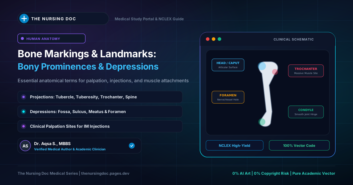
Classification Architecture of Human Bone Markings
Topographical taxonomy: Articular joints, muscle attachment projections, and neurovascular openings
Introduction: What are Bony Markings?
The surface of a dried human bone is rarely smooth. Instead, it presents various distinct bony markings in the form of roughened areas, elevated processes, shallow depressions, holes (apertures), and elongated grooves.
In clinical medicine, surgery, and nursing practice, these anatomical markings are not merely academic terminology—they serve as crucial surface landmarks. Nurses routinely palpate these landmarks to safely locate intramuscular (IM) injection sites (e.g., greater trochanter for ventrogluteal injections), gauge peripheral pulse points, determine landmark levels for lumbar punctures, and assess for bone fractures and localized edema.
Articular Surfaces Definitions
Articular surfaces comprise the specific portions of bones that interface directly with adjacent bones to construct synovial or cartilaginous joints. Because they experience high friction and mechanical compression, they are typically capped in living tissue with smooth, low-friction hyaline cartilage.
🔘 Head (Caput)
A large, rounded, hemispherical articular prominence, typically supported by and set off from the rest of the shaft by a constricted segment termed the neck.
⚪ Capitulum
A small, rounded, dome-shaped articular eminence (derived from the Latin word meaning "small head").
🪢 Trochlea
A pulley-shaped articular surface featuring a central groove bounded by elevated medial and lateral margins.
🤜 Condyle
A large, rounded protuberance or knuckle-like projection located at the terminal articular end of a bone, typically occurring in pairs (medial and lateral).
💎 Facet
A small, smooth, flat or slightly curved articular gliding surface.
🥣 Fossa
A hollow, broad, depressed region on a bone that accommodates an opposing bony process during joint articulation or cradles soft organ tissue.
Figure 1: Distal Humerus Articular Architecture
Comparing the rounded lateral capitulum, pulley trochlea, and reception coronoid fossa
Non-Articular Surfaces (Projections & Ridges)
Non-articular bone markings do not form joints. Instead, they serve primarily as mechanical anchor points for skeletal muscles, dense tendons, and stabilizing ligaments. The size and roughness of these markings are directly proportional to the muscular force exerted upon them throughout life.
1. Process
A broad descriptive term denoting any prominent projection or outgrowth extending from a bone, frequently named after its anatomical morphology:
- Coracoid process of the scapula: Resembles the curved beak of a crow (coracoid = crow-like).
- Mastoid process: A blunt, rounded conical projection of the temporal bone meaning "breast-shaped".
- Styloid process: A long, slender, sharply pointed projection of the temporal bone shaped like a stylus (an ancient writing pen).
2. Trochanter
A very large, blunt, irregularly shaped prominence found exclusively on the proximal femur.
💡 Examples: Greater trochanter (key landmark for hip palpation and gluteus medius insertion) and lesser trochanter (iliopsoas insertion) of the femur.
3. Tubercle vs. Tuberosity
4. Malleolus
A distinctive bony projection shaped like a hammerhead located at the terminal distal ends of the lower leg bones.
💡 Examples: Medial malleolus of the tibia (inner ankle bone); Lateral malleolus of the fibula (outer ankle bone).
5. Epicondyle
An elevated projection situated on or immediately above an articular condyle, serving as a non-articular anchor for collateral ligaments and forearm/leg muscles.
💡 Examples: Lateral and medial epicondyles of the humerus; lateral and medial epicondyles of the femur.
6. Spine
A sharp, slender, or short pointed thorn-like projection from a bone.
💡 Examples: Anterior nasal spine of the maxilla; the prominent spine of the scapula on its dorsal posterior surface; ischial spine of the pelvis.
7. Hamulus
A specialized hook-like or claw-shaped curved projection extending from a bone.
💡 Example: Pterygoid hamulus located at the inferior terminus of the medial pterygoid plate of the sphenoid bone (pivots the tensor veli palatini tendon).
8. Ridge, Crest, & Line (Linear Elevations)
Linear elevations are graded by their prominence and sharpness:
9. Fovea & Notch
Grooves and Holes (Apertures & Passageways)
Bones must allow blood vessels to enter for marrow nourishment and provide safe passageways for peripheral and cranial nerves traversing between body compartments. These pathways are categorized as under:
⚡ Fissure
A narrow, slit-like cleft or groove traversing through or between adjacent bones.
🕳️ Foramen (Plural: Foramina)
A round or oval hole, perforation, or opening that allows vessels or nerves to pass directly into or through a bone.
🚇 Meatus
A short, tube-like canal or passage extending completely through the interior substance of a bone.
〰️ Sulcus (Groove)
A shallow elongated groove, furrow, or channel carved on the external surface of a bone to house a tendon or vessel without compressing it.
🚪 Hiatus
A cleft, gap, naturally occurring opening, or deficiency in a bone surface meant for the physiological passage of nerves, vessels, or therapeutic access.
Bedside Nursing Application: Crucial Bony Landmarks
In clinical nursing exams (NCLEX) and hospital wards, safe medication administration and physical assessments depend entirely on your ability to palpate bony landmarks:
Place your palm over the patient's greater trochanter, index finger on the anterior superior iliac spine (ASIS), and middle finger along the iliac crest. The resulting "V" triangle is the safest deep gluteal injection site, completely avoiding the sciatic nerve.
The medial malleolus is the landmark for palpating the posterior tibial artery pulse (just posterior and inferior to the bone). Both malleoli are standard sites for evaluating peripheral pitting edema depth (+1 to +4 scale).
A horizontal line drawn across the highest points of both iliac crests crosses the L4 spinous process (Tuffier's line). This guides the needle safely into the L3-L4 or L4-L5 interspace below the termination of the adult spinal cord (conus medullaris at L1-L2).
Quick-Review Summary: Bony Markings Reference Table
| Landmark Type | Primary Category | Structural Definition | High-Yield Clinical Example |
|---|---|---|---|
| Head | Articular | Large rounded articular ball on a constricted neck | Head of Femur |
| Capitulum | Articular | Small, rounded articular eminence ("little head") | Distal lateral humerus |
| Trochlea | Articular | Pulley-shaped articular surface with groove | Distal medial humerus (elbow hinge) |
| Condyle | Articular | Rounded paired protuberance at terminal joint end | Femoral condyles (knee joint) |
| Trochanter | Projection | Very large, blunt prominence (femur only) | Greater & Lesser trochanters of femur |
| Tuberosity | Projection | Rough, elevated patch for tendon attachment | Deltoid tuberosity of humerus |
| Malleolus | Projection | Hammerhead-shaped ankle projection | Medial (tibia) & Lateral (fibula) |
| Epicondyle | Projection | Non-articular projection situated above a condyle | Medial & lateral epicondyles of humerus |
| Foramen | Aperture | Hole or opening through bone for nerves/vessels | Nutrient foramen, Foramen magnum |
| Meatus | Aperture | Canal or tube-like passage within bone | External auditory meatus |
| Sulcus | Groove | Shallow groove or furrow accommodating tendon | Intertubercular sulcus (bicipital groove) |
| Hiatus | Aperture | Gap or deficient opening in bone surface | Sacral hiatus (caudal anesthesia) |
Curated and medically verified by Dr. Aqsa S. for nursing students.
← Back to All Study Notes