Cardiovascular Circulation & Cardiac Dynamics: Systemic vs. Pulmonary Circuits & Wiggers Cycle
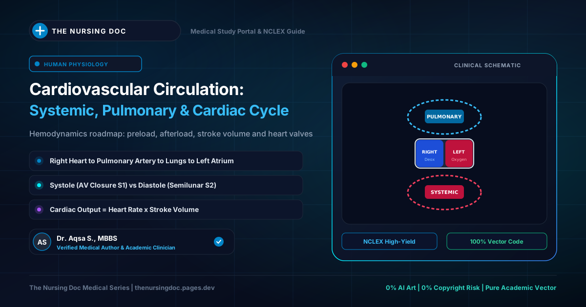
The Dual Cardiovascular Circulatory Loop: Systemic vs. Pulmonary
Topographical schematic of pulmonary gas exchange and systemic peripheral tissue perfusion
1. What is Circulation? The Dual Circulatory Pathways
Circulation represents the continuous hydrodynamic transport system responsible for circulating blood and interstitial lymph throughout every tissue plane of the human body. Physiologically, cardiovascular blood flow is divided into two distinct, serially linked circulatory loops:
🩸 A. Systemic Circulation (Peripheral / General)
Also termed the general or peripheral circulation, this extensive circuit supplies oxygen, glucose, hormones, and immune cells to all active tissues of the body except the lungs.
Left Ventricle ➔ Aorta ➔ Muscular Arteries ➔ Arterioles ➔ Capillaries ➔ Venules ➔ Veins ➔ Superior & Inferior Vena Cava ➔ Right Atrium.
🫁 B. Pulmonary Circulation (Lesser Circuit)
This dedicated low-resistance vascular loop conveys venous deoxygenated blood from the right heart into the pulmonary microvascular bed for carbon dioxide clearance and alveolar oxygen saturation.
Right Ventricle ➔ Pulmonary Trunk & Arteries ➔ Pulmonary Arterioles ➔ Pulmonary Capillaries ➔ Pulmonary Venules & Veins ➔ Left Atrium.
Functional Anatomy of the Vascular Tree
The cardiovascular circuit is engineered with specialized anatomical conduits, each adapted for distinct hemodynamic roles:
🫀 The Heart (Central Muscular Pump)
A conical, hollow, four-chambered muscular organ situated obliquely in the middle mediastinum of the thoracic cavity, enveloped and lubricated within the fibroserous pericardium. It operates as dual synchronized pumps: the right heart drives pulmonary flow, while the thicker left heart generates systemic pressure.
🔴 Arteries (Conduit Vessels)
Thick, high-pressure vessels that convey blood away from the heart. Their walls are constructed of three distinct histological tunics: Tunica Intima (endothelium), Tunica Media (rich smooth muscle and elastic elastin fibers), and Tunica Adventitia (fibrous connective tissue sheath).
🎛️ Arterioles (Resistance Regulators)
The smallest terminal branches of arteries that deliver blood into the capillary beds. Their heavily innervated tunica media smooth muscle allows dynamic vasoconstriction and vasodilation. By adjusting luminal caliber, arterioles represent the primary site of Systemic Vascular Resistance (SVR) and govern arterial blood pressure ($BP = CO \times SVR$).
🕸️ Capillaries (Microvascular Exchange Sites)
Diffuse microscopic networks connecting arterioles and venules. Constructed of only a solitary layer of endothelial cells and a basal lamina, capillaries lack tunica media and adventitia, facilitating rapid trans-membrane diffusion of gases, electrolytes, and nutrients.
Capillary Density: Varies proportionally with tissue metabolic demand: extraordinarily rich in skeletal muscle, liver, renal cortex, lungs, and the central nervous system; sparse in dense tendons and absent in cartilage and cornea.
🔵 Venules & Veins (Capacitance Reservoirs)
Venules collect post-capillary blood and coalesce into veins, which return blood back to the heart. Veins possess the same three tunics as arteries, but have significantly thinner tunica media, wider lumens, and higher compliance. They house approximately 60% to 70% of total circulating blood volume (the venous capacitance reservoir) and feature one-way endothelial semilunar valves preventing retrograde flow against gravity.
Properties of Cardiac Muscle & Conduction
Cardiac muscle (myocardium) is involuntary in function and cross-striated in microscopic appearance, possessing unique electrophysiological attributes:
Figure 1: Specialized Electrical Conduction Pathway of the Heart
Spontaneous automaticity from the SA node propagating to ventricular Purkinje fibers
- Absolute Refractory Period (ARP): Zero excitability regardless of stimulus strength.
- Relative Refractory Period (RRP): Muscle can respond only to a supranormal stimulus.
The Cardiac Cycle: Wiggers 8-Stage Timeline (0.8 Seconds)
The Cardiac Cycle encompasses all physiological events occurring from the beginning of one heartbeat to the onset of the next. At a normal resting heart rate of 75 beats per minute, each cardiac cycle is completed in precisely 0.8 seconds.
✊ A. Ventricular Systole (Total: 0.303 sec)
Period of active ventricular contraction and blood ejection:
🫀 B. Ventricular Diastole (Total: 0.495 sec)
Period of ventricular relaxation, repolarization, and ventricular filling:
Heart Sounds: Auscultation, Valves, & Murmurs
Heart sounds are auscultated using a stethoscope (invented in 1816 by French physician René Laennec). They are generated by the physical closure of heart valves and hemodynamic turbulence within chambers:
1st Heart Sound (S₁): "LUB"
Duration: 0.15 secProduced at the very onset of ventricular systole by the simultaneous closure of the Atrioventricular (Tricuspid & Mitral) valves.
- ECG Correlation: Follows immediately after the QRS complex.
- Best Heard: Loudest at the apex of the heart (5th intercostal space, midclavicular line).
- Pitch: Lower pitch, softer, but longer in duration.
2nd Heart Sound (S₂): "DUB"
Duration: 0.12 secProduced at the end of ventricular systole / onset of diastole by the closure of the Semilunar (Aortic & Pulmonary) valves.
- ECG Correlation: Corresponds with the end of the T wave.
- Best Heard: Loudest at the base of the heart (2nd intercostal spaces).
- Pitch: Higher pitch, sharper, and shorter in duration.
🔊 Cardiac Murmurs (Turbulent Flow)
Murmurs are abnormal whooshing or blowing sounds generated by turbulent blood flow. They arise from diseased, stenotic valves (narrowed opening causing a jet), regurgitant valves (incompetent leaflets causing backflow leakage), or congenital septal wall defects (ASD, VSD).
Bedside Nursing Application & Cardiac Auscultation Pearls
Cardiovascular circulation and valve timings dictate bedside physical exams and acute monitoring:
• A: Aortic (2nd R ICS)
• P: Pulmonic (2nd L ICS)
• E: Erb's Point (3rd L ICS - S1/S2 equal)
• T: Tricuspid (4th L ICS sternal border)
• M: Mitral/Apex (5th L ICS midclavicular line)
Heard immediately after S2 ("Ken-TUCK-y"). Caused by rapid, turbulent inflow hitting a dilated, volume-overloaded ventricle in congestive heart failure. Always an abnormal finding in adults over age 40.
Because arterioles regulate 80% of SVR, IV vasodilators (Hydralazine, Nitroprusside) dilate arteriolar smooth muscle to rapidly plunge systemic vascular resistance, unloading the left ventricle in hypertensive crisis.
Quick-Review Summary: Cardiovascular System Comparison
| Circulatory Parameter | Origin Chamber | Primary Destination | Pressure Profile | Key Functional Role |
|---|---|---|---|---|
| Systemic Circuit | Left Ventricle | All body tissues & organs | High Pressure (~120/80 mmHg) | Perfusion, nutrient delivery, waste pickup |
| Pulmonary Circuit | Right Ventricle | Pulmonary alveolar capillaries | Low Pressure (~25/10 mmHg) | CO₂ clearance & blood re-oxygenation |
| Arterioles | Muscular Arteries | Capillary bed microcirculation | Variable (smooth muscle tone) | Controls Systemic Vascular Resistance (SVR) |
| S₁ Heart Sound ("Lub") | Onset of Systole | Closure of AV (Mitral/Tricuspid) valves | Isovolumetric ventricular spike | Prevents retrograde backflow into atria |
| S₂ Heart Sound ("Dub") | End of Systole | Closure of Semilunar (Aortic/Pulm) valves | Isovolumetric ventricular drop | Prevents arterial backflow into ventricles |
Curated and medically verified by Dr. Aqsa S. for nursing students.
← Back to All Study Notes