Anemia Simplified: Types, Etiological Classifications, Aplastic Anemia, & Megaloblastic Disorders
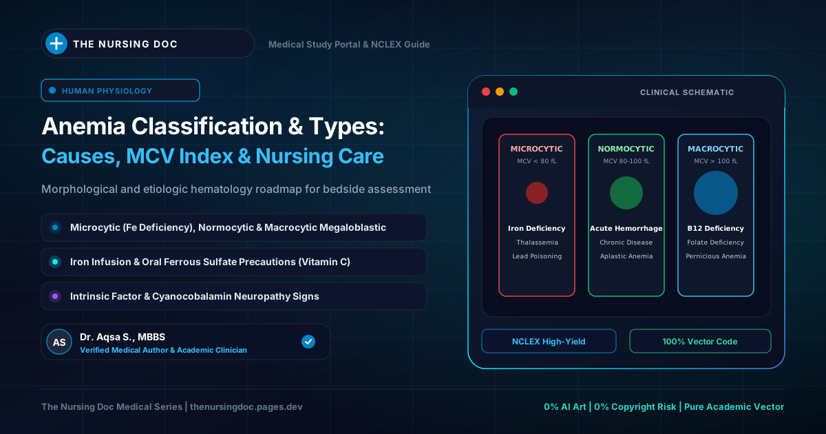
Etiological Classification & Pathophysiological Pathways of Anemia
The 3 Core Mechanisms: Blood Loss, Hemolysis, and Defective Red Cell Production
1. Definition of Anemia
Anemia is defined as a decreased concentration of hemoglobin (Hb%) in the circulating blood below the normal reference range established for the age, biological sex, and physiological state (e.g., pregnancy) of the individual:
2. Comprehensive Classification of Anemia
From an etiological standpoint, anemia arises through three fundamental pathophysiological disturbances:
A) Blood Loss Anemia
- Acute Post-Hemorrhagic Anemia: Sudden, massive blood volume loss resulting from major mechanical trauma, vascular rupture, or surgical catastrophe.
-
Chronic Hemorrhagic Anemia: Occult or low-grade persistent blood loss that insidiously depletes body iron reserves over weeks or months. Common causes include:
- Peptic Ulcer Disease (gastric and duodenal ulcers)
- Parasitic intestinal hookworm infestation (Ancylostoma duodenale, Necator americanus)
- Gastrointestinal or genitourinary malignancy (e.g., colon carcinoma)
- Underlying bleeding disorders (von Willebrand disease, hemophilia, chronic menorrhagia)
B) Increased Rate of RBC Destruction (Hemolytic Anemia)
In hemolytic states, the red blood cell lifespan falls significantly below the standard 120-day physiological benchmark. The bone marrow attempts compensation by increasing reticulocyte output by 6–8 fold, but severe anemia develops when the rate of destruction outpaces compensatory erythropoiesis.
C) Impaired Red Cell Production (Hypoproliferative & Maturation Disorders)
-
1. Disturbance of Proliferation and Differentiation of Pluripotent Stem Cells:
- Aplastic Anemia (idiopathic or secondary marrow failure)
- Pure Red Cell Aplasia (PRCA)
- Anemia of Chronic Kidney Disease (deficient renal erythropoietin production)
- Anemia due to endocrine disorders (hypothyroidism, hypopituitarism)
-
2. Disturbance of Proliferation and Maturation of RBCs:
- Defective DNA Synthesis: Deficiency or impaired utilization of Vitamin B12 and Folic Acid (Megaloblastic Anemia).
- Defective Hemoglobin Synthesis:
- Deficient Heme synthesis: Iron Deficiency Anemia.
- Deficient Globin chain synthesis: Thalassemia (Alpha and Beta).
- Abnormal Hemoglobin Structure: Sickle Cell Anemia (point mutation in beta-globin substituting valine for glutamic acid at codon 6).
3. Aplastic Anemia (Bone Marrow Aplasia)
In Aplastic Anemia, the circulating count of all three major peripheral blood cell lines—erythrocytes, leukocytes, and thrombocytes—is profoundly diminished. This condition of generalized marrow failure is clinically termed Pancytopenia.
The underlying pathophysiology is bone marrow aplasia (congenital hypoplasia, physical absence, or toxic destruction of multipotent hematopoietic stem cells), resulting in marrow replacement by inactive adipose tissue.
Etiological Classification of Aplastic Anemia:
- Congenital: Fanconi's Anemia (autosomal recessive DNA-repair defect associated with skeletal anomalies and café-au-lait spots).
- Acquired: Secondary autoimmune T-cell-mediated destruction of hematopoietic stem cells.
- Radiation: Exposure to ionizing radiation hazards, atomic blasts, or occupational industrial X-rays.
- Chemical Toxins: Benzene, volatile organic solvents, insecticides, and petrochemical compounds.
- Pharmacological Drugs: Chloramphenicol (dose-dependent and idiosyncratic marrow shutdown), Sulphonamides, Phenylbutazone, Chlorpheniramine, Chlorpromazine, Carbimazole, Methotrexate, Gold salts.
- Miscellaneous Factors: Pregnancy, disseminated Tuberculosis (miliary TB), viral infections (EBV, Hepatitis, Parvovirus B19), and severe socio-economic deprivation.
Clinical Manifestations of Aplastic Anemia
- Progressive Anemia: Profound lethargy, pallor of the skin and conjunctivae, fatigue, and exertional breathlessness due to decreased red blood cell mass.
- Hemorrhagic Manifestations: Petechial hemorrhages in the dermis and mucous membranes, ecchymosis, spontaneous bleeding from the gums (gingival bleeding), epistaxis (nosebleeds), and heavy menorrhagia directly resulting from severe thrombocytopenia.
- Infectious Manifestations: High spiking fevers, bacterial pharyngitis, and necrotic ulcerations in the oral cavity and throat due to severe granulocytopenia (neutrophil depletion).
Treatment Protocol
- Bone Marrow Transplantation (BMT): Allogeneic hematopoietic stem cell transplantation remains the definitive curative modality, particularly in young patients with HLA-matched sibling donors.
- Transfusion Support: Regular transfusions of packed red blood cells (PRBCs) to maintain hemodynamic oxygen delivery, and platelet concentrates to avert catastrophic intracranial or visceral hemorrhage.
- Immunosuppressive Therapy: Antithymocyte Globulin (ATG), Cyclosporine, and high-dose corticosteroid regimens to halt autoimmune stem cell destruction in non-transplant candidates.
- Infection Control: Broad-spectrum empirical intravenous antibiotics and antifungal prophylaxis in a protective reverse-isolation environment.
4. Megaloblastic Anemia
Megaloblastic Anemia represents a distinct subgroup of macrocytic anemias characterized by the presence of large, abnormal, oval-shaped erythroblasts (megaloblasts) within the bone marrow and hypersegmented neutrophils in the peripheral circulation.
The fundamental biochemical defect is impaired nuclear DNA synthesis with preserved cytoplasmic RNA and protein synthesis (nuclear-cytoplasmic dyssynchrony). There are two principal etiologies:
- Vitamin B12 (Cobalamin) Deficiency
- Folic Acid (Folate) Deficiency
Etiology of Vitamin B12 Deficiency
- Inadequate Dietary Intake: Strict long-term vegans who consume zero animal proteins, dairy products, or eggs (Vitamin B12 is synthesized exclusively by microorganisms and found solely in animal products).
-
Impaired Intestinal Absorption:
- Intrinsic Factor (IF) Deficiency: Pernicious Anemia (autoimmune destruction of gastric parietal cells), Total or Partial Gastrectomy, and congenital intrinsic factor absence.
- Disorders of the Terminal Ileum: Tropical sprue, Celiac disease (nontropical sprue), Crohn's disease (regional enteritis), surgical ileal resection, lymphoma, and granulomatous enteritis.
-
Intraluminal Competition for Vitamin B12:
- Fish tapeworm infestation (Diphyllobothrium latum, which consumes up to 80% of dietary B12).
- Bacterial Blind Loop Syndrome (jejunal diverticulosis or surgical loops with bacterial overgrowth that metabolize cobalamin).
Etiology of Folic Acid Deficiency
-
Inadequate Dietary Intake:
- Unbalanced diet lacking raw green leafy vegetables, citrus fruits, and liver.
- Excessive Cooking / Boiling: Folate is heat-labile and destroyed by prolonged boiling.
- Chronic alcoholics (alcohol suppresses folate absorption and hepatic release).
- Elderly individuals living alone, severe poverty, and starvation diets.
-
Malabsorption Syndromes & Drug Interactions:
- Tropical sprue and gluten-sensitive enteropathy.
- Anticonvulsant medications (Diphenylhydantoin / Phenytoin, barbiturates), which inhibit intestinal folate conjugase.
-
Increased Physiological / Pathological Demands:
- Pregnancy (fetal growth requires 5–10x higher folate; deficiency triggers Neural Tube Defects).
- Lactation and rapid growth in infancy.
- Widespread malignancy and chronic hemolytic states with rapid cell turnover.
- Antineoplastic Drugs Impairing DNA Metabolism: Methotrexate (dihydrofolate reductase inhibitor), 6-Mercaptopurine, 5-Fluorouracil (5-FU), and Cytosine arabinoside.
5. Pernicious Anemia
Pernicious Anemia is a specific autoimmune form of megaloblastic anemia caused by chronic autoimmune atrophic gastritis. Progressive destruction of gastric oxyntic (parietal) cells leads to severe achlorhydria (absence of gastric acid) and complete failure of Intrinsic Factor (IF) secretion.
Without Intrinsic Factor, Vitamin B12 cannot bind or be recognized by the cubam receptor complex in the terminal ileum, rendering oral cobalamin completely unabsorbable.
Clinical Features of Pernicious Anemia
Patients suffering from pernicious anemia typically present with an insidious, gradual clinical course:
- Systemic Symptoms: Progressive weakness, unintentional weight loss, easy fatigability, dyspnea on exertion, lightheadedness, and syncope.
- Dermal & Mucosal Signs: Distinctive lemon-yellow skin pallor (combination of pallor and mild indirect hyperbilirubinemia from intramedullary hemolysis).
- Oral Signs (Hunter's Glossitis): The tongue appears beefy red, painful, and inflamed in acute stages, progressing to a classically smooth, shiny, atrophic tongue due to loss of lingual papillae.
- Abdominal Signs: Hepatosplenomegaly (mild splenomegaly is palpable in approximately 10–15% of cases).
Medical Management Protocol
Because the oral route is permanently compromised by intrinsic factor absence, treatment requires parenteral administration:
• Loading Phase: Hydroxocobalamin 1,000 micrograms (mcg) intramuscularly (IM) every other day or twice weekly to a total cumulative dose of 4,000 – 6,000 mcg over 3 weeks.
• Maintenance Phase: 1,000 micrograms IM every 3 months for the rest of the patient's life.
6. Folic Acid Deficiency Anemia
Folic acid deficiency produces a megaloblastic blood picture indistinguishable from Vitamin B12 deficiency on peripheral smear (both exhibit macro-ovalocytes with MCV > 100 fL and hypersegmented neutrophils with ≥ 6 lobes).
Clinical Presentation
Mild folate deficiency may remain subclinical, but severe deficiency presents with severe malnutrition, pronounced weight loss, weakness, sleep disturbances (insomnia), impaired cognitive concentration, painful glossitis, and cheilosis / angular cheilitis (crusting fissures and ulceration at the labial commissures).
🚨 Critical Clinical Safety Rule: The Folate Neurological Trap
REMEMBER: In stark contrast to Vitamin B12 deficiency, neurological abnormalities DO NOT OCCUR in pure folic acid deficiency.
However, Folic acid must NEVER be administered alone in a patient with undiagnosed or suspected Vitamin B12 depletion! High-dose folic acid drives the DNA synthesis pathway, partially clearing the hematological anemia, while the uncorrected cobalamin deficiency silently accelerates irreversible demyelination of the posterior and lateral spinal cord columns (Subacute Combined Degeneration) resulting in permanent spastic paraplegia!
Therapeutic Protocol for Folate Deficiency
- Active Deficiency Dosing: Oral Folic acid 5 mg daily for 4 months (sufficient to replace exhausted body reserves).
- Prophylactic Indications:
- Pre-conception and Pregnancy: 400 mcg daily (4–5 mg daily in high-risk mothers) to prevent neural tube defects (spina bifida, anencephaly).
- Chronic Hemolytic Anemias: Lifelong prophylaxis in Thalassemia major and Sickle Cell Disease to meet excessive marrow turnover demands.
7. High-Yield Comparative Summary Table
| Parameter | Aplastic Anemia | Pernicious Anemia (B12) | Folate Deficiency |
|---|---|---|---|
| Primary Mechanism | Bone Marrow Stem Cell Aplasia | Autoimmune Intrinsic Factor loss | Dietary deficiency / Malabsorption |
| Peripheral Smear | Pancytopenia (Normocytic) | Megaloblasts, Macrocytic (MCV > 100) | Megaloblasts, Macrocytic (MCV > 100) |
| Neurological Signs | Absent | Present (Paresthesias, Ataxia, SCD) | Strictly Absent |
| Distinctive Signs | Petechiae, epistaxis, mouth ulcers | Smooth atrophic tongue, lemon pallor | Cheilosis, severe malnutrition |
| Definitive Therapy | BMT, transfusions, immunosuppressants | Hydroxocobalamin 1000 mcg IM for life | Oral Folic acid 5 mg daily |
📝 NCLEX High-Yield Pearls:
- Pancytopenia = Bone Marrow Biopsy Needed: When a CBC shows low RBCs, low WBCs, and low Platelets, think Aplastic Anemia or acute leukemia. Put the patient on Bleeding Precautions (soft toothbrush, no IM injections) and Neutropenic Isolation.
- The B12 "Neuro" Rule: B12 deficiency affects the brain and nerves (Cobalamin myelinates axons). Folate deficiency does NOT cause peripheral neuropathy.
- Pernicious Anemia Is Lifelong: Patient education must emphasize that IM cobalamin cannot be stopped when symptoms resolve—it is required for life.
Written & Medically Reviewed by Dr. Aqsa S.
Medical Doctor specializing in clinical hematology, physiology education, and exam preparation. Dedicated to translating complex clinical pathology into high-yield, life-saving clarity for nursing students.