Blood Physiology: Plasma Composition, Erythrocyte Morphology, & Erythropoiesis Guide
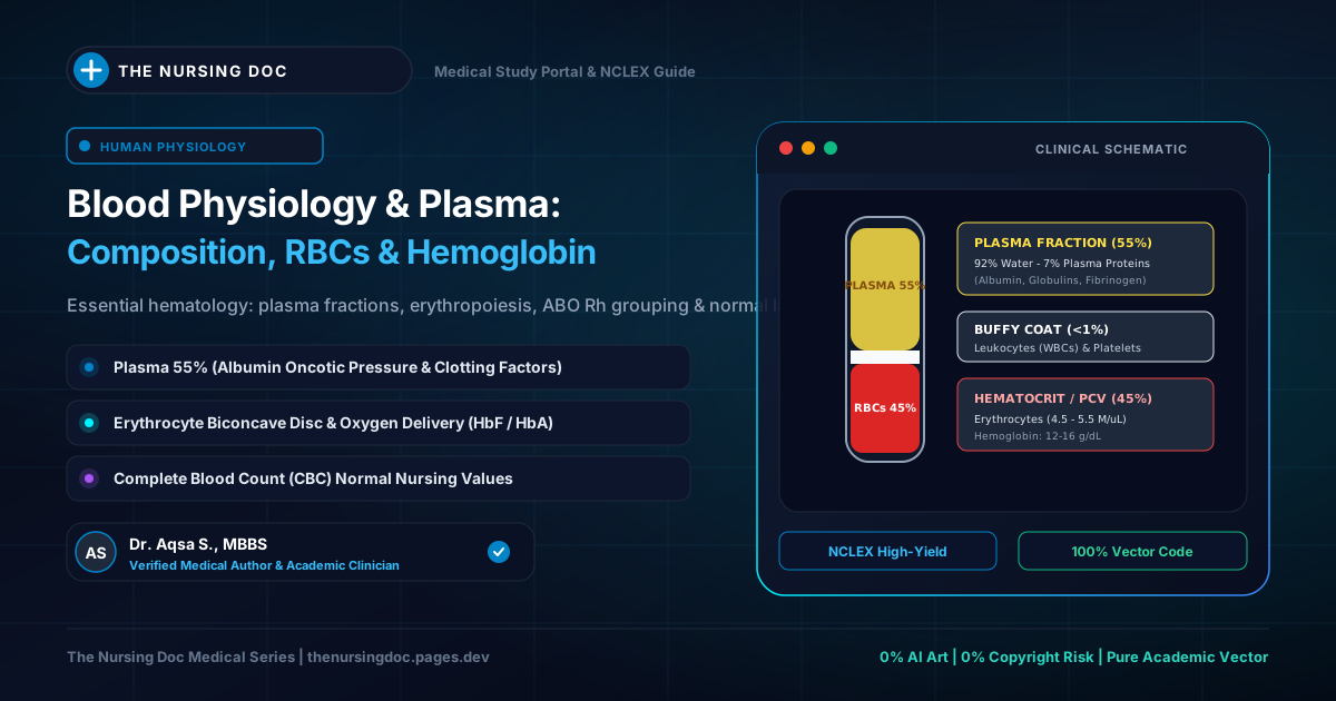
⚡ High-Yield Clinical Summary
- Embryological Origin: Blood is a specialized fluid connective tissue derived from the embryonic mesoderm.
- Two Major Fractions: Cellular formed elements (45%: RBCs, WBCs, Platelets) and non-cellular liquid Plasma (55%: 91% water, 7-8% proteins, 1-2% solutes).
- Physical Parameters: Adult blood volume is ~5 Liters (4.5 L in females, 450 mL in neonates), slightly alkaline with a tightly controlled pH of 7.4 (7.35–7.45), and 5 times more viscous than water.
- Mature RBC Features: Non-nucleated biconcave disks devoid of mitochondria; rely exclusively on anaerobic glycolysis for ATP; average lifespan is 120 days (70–90 days in neonates).
- Erythropoiesis Sequence: 1st trimester (Yolk sac) → 2nd trimester (Liver & Spleen) → 3rd trimester & Adult (Red Bone Marrow: ribs, sternum, vertebrae, ilium). Controlled by renal Erythropoietin (EPO) in response to tissue hypoxia.
Human Blood Tissue: Architecture, Fractions, & Formed Elements
Complete structural map of circulating cardiovascular fluid
1. Definition & Primary Composition of Blood
In clinical physiology, blood is classified as a specialized fluid connective tissue of mesodermal origin. Circulating continuously through the cardiovascular system, blood serves as the primary convective transport medium delivering oxygen, nutrients, electrolytes, and hormones to peripheral tissues while retrieving metabolic end-products and carbon dioxide.
When whole blood is anticoagulated and centrifuged, it separates into two distinct fractions:
Cellular Part (Formed Elements)
- 1. Erythrocytes (Red Blood Cells / RBCs): Gas-transporting cells carrying hemoglobin (Hb).
- 2. Leukocytes (White Blood Cells / WBCs): Immune defenders combating microbial invasion.
- 3. Thrombocytes (Platelets): Anucleated cytoplasmic fragments crucial for primary hemostasis and clot formation.
Non-Cellular Part (Blood Plasma)
The straw-colored extracellular fluid vehicle of blood. Composed of 91% water and 9% solids/dissolved solutes (proteins, electrolytes, nutrients, metabolic wastes, and regulatory enzymes).
Figure 1: Fractionation of Whole Blood by Centrifugation (Hematocrit)
Visual representation of fluid plasma vs. cellular formed elements
Detailed Chemical Breakdown of Blood Plasma
The 9% solid matter of plasma comprises a vast array of vital organic and inorganic biochemical constituents:
| Chemical Class | Constituents & Concentrations | Primary Physiological Role |
|---|---|---|
| Plasma Proteins (7–8%) |
• Albumin (4.5 – 5.5%): Most abundant • Globulins (1.3 – 2.0%): Alpha, Beta, Gamma • Fibrinogen (0.2%): Soluble clotting factor • Prothrombin (0.1%): Coagulation precursor • Complement System: ~20 enzymatic proteins |
Generates intravascular colloid osmotic (oncotic) pressure (~25 mmHg via albumin), immunity (gamma globulins/antibodies), and enzymatic blood coagulation. |
| Non-Protein Nitrogenous (NPN) Substances | Urea, Uric acid, Creatinine, Free Amino acids, Ammonia. | Metabolic waste products filtered by renal glomeruli; clinical indicators of kidney excretory performance. |
| Non-Nitrogenous Organic Solutes | Glucose, Cholesterol, Galactose, Phospholipids, Triglycerides. | Primary energy substrates and structural lipid carriers traversing between liver and peripheral cells. |
| Regulatory Enzymes & Pigments | Amylase, Carbonic Anhydrase, Lipase, Phosphatase, SGPT (ALT), SGOT (AST), LDH; Pigment: Bilirubin. | Catalyze metabolic conversions and acid-base buffering; Bilirubin represents the normal degradation breakdown product of heme catabolism. |
| Inorganic Electrolytes | Sodium (Na⁺), Potassium (K⁺), Chloride (Cl⁻), Calcium (Ca²⁺), Bicarbonate (HCO₃⁻), Iodine, Magnesium (Mg²⁺), Phosphate. | Govern neuromuscular excitability, plasma osmolarity (285–295 mOsm/kg), and systemic acid-base equilibrium. |
2. Physical & Chemical Properties of Blood
Nurses and clinicians routinely evaluate the fundamental physical parameters of circulating blood to identify acute pathological states:
- • Color: Blood is naturally red due to iron-containing hemoglobin. Highly oxygenated arterial blood (rich in Oxyhemoglobin) appears bright scarlet red, whereas deoxygenated venous blood (Deoxyhemoglobin) exhibits a darker, purple-reddish hue.
- • Volume: The average adult systemic blood volume is approximately 5 Liters (accounting for ~7-8% of total adult body weight). In adult females, average volume is slightly lower at 4.5 Liters. In newborn infants, total blood volume is only approximately 450 mL (~80-85 mL/kg).
- • pH: Blood is slightly alkaline. The physiological set-point of arterial blood pH in normal resting conditions is tightly maintained at 7.40 (strictly buffered within the range of 7.35 to 7.45). Values < 7.35 define acidemia; values > 7.45 define alkalemia.
- • Viscosity: Whole blood is approximately 5 times more viscous than pure water. This internal fluid friction is predominantly generated by the physical concentration of circulating red blood cells (hematocrit) and high-molecular-weight plasma proteins (fibrinogen and globulins).
3. Red Blood Cells (Erythrocytes): Morphology & Architecture
Erythrocytes (RBCs) are the most numerous cells in human blood. They are specialized, non-nucleated, circular biconcave discs dedicated to transporting respiratory gases (O₂ and CO₂) between pulmonary alveoli and systemic capillary beds.
Normal Physiological RBC Counts
Internal Composition of RBCs
During terminal maturation in the bone marrow, the erythrocyte expels its nucleus, mitochondria, ribosomes, and Golgi apparatus to maximize cytoplasmic storage space for Hemoglobin (Hb):
- Water: 65% of total erythrocyte mass.
- Solids & Semisolids: 35% of erythrocyte mass.
- Hemoglobin (Hb): Accounts for 33% of the total RBC weight and over 90% of its dry solid mass.
- Organic & Inorganic Substances (2%): Structural membrane proteins (spectrin, ankyrin), phospholipids, cholesterol, urea, creatinine, free amino acids, and vital electrolytes (potassium, chloride, bicarbonate).
Biconcave Disk Morphology: Fluid Dynamics & Physiology
Erythrocytes are shaped as circular biconcave disks (mean diameter ~7.2–7.8 µm, thickness 2.5 µm at the peripheral rim and <1.0 µm at the central pallor), exhibiting a dumbbell-shaped cross-section and a torus-shaped peripheral rim.
Why Did Nature Select the Biconcave Shape?
- Surface Area to Volume Ratio: Maximizes the surface area available for instantaneous diffusion of O₂ and CO₂ across the plasma membrane.
- Laminar Flow Optimization: Optimizes hydrodynamic flow properties through large arteries, maximizing smooth central laminar streaming while minimizing platelet scatter toward endothelial walls (reducing spontaneous thrombogenesis).
- Extreme Deformability: With a flexible spectrin-actin cytoskeleton, the erythrocyte flexes and folds upon itself to traverse microscopic capillaries measuring only 4 to 5 µm in caliber without rupturing.
Figure 2: Geometric Dimensions & Cross-Section of a Mature Human Erythrocyte
Top-down surface view vs. transverse dumbbell cross-section
Lifespan & Energy Metabolism
The average physiological lifespan of an adult circulating erythrocyte is 120 days. In neonates, RBCs turn over significantly faster, displaying a reduced lifespan of 70 to 90 days.
Because mature RBCs completely lack mitochondria, they cannot execute the citric acid (Krebs) cycle or oxidative phosphorylation. Instead, erythrocytes depend strictly upon plasma glucose to generate ATP through anaerobic glycolysis (Embden-Meyerhof pathway). This ATP powers active cation pumps (Na⁺/K⁺-ATPase) that maintain intracellular osmotic balance and preserve biconcave membrane integrity.
4. Erythropoiesis: Stages, Sites, & Regulatory Factors
Erythropoiesis is the physiological process of erythrocyte genesis and maturation. The anatomical site of RBC production shifts systematically across developmental stages:
1st Trimester of Fetal Development (0 – 3 Months): Mesoblastic Stage
Erythrocytes are generated extra-embryonically within the blood islands of the fetal yolk sac.
2nd Trimester of Fetal Development (3 – 6 Months): Hepatic & Splenic Stage
Erythropoiesis migrates into the embryonic liver (predominant organ), with secondary contributions from the spleen and lymphoid tissue.
3rd Trimester & Postnatal Life: Myeloid (Bone Marrow) Stage
From the 7th fetal month onward, the red bone marrow becomes the primary lifelong hematopoietic factory:
- Birth up to Age 5: Red marrow in practically all bones actively manufactures red blood cells.
- Age 5 to 20: Shafts (diaphyses) of long tubular bones gradually undergo fatty metamorphosis into inactive yellow marrow.
- After Age 20 to 25: Red blood cell production becomes confined exclusively to the red marrow of membranous and flat bones (e.g., Sternum, Ribs, Vertebral bodies, Ilium/Pelvis, Skull, and proximal epiphyses of femur/humerus).
Essential Nutritional & Biochemical Factors for Erythropoiesis
Successful maturation of erythrocytes from pluripotent stem cells (CFU-E) requires several mandatory cofactors:
- 1. Vitamin B12 (Cyanocobalamin) & Folic Acid: Indispensable for DNA thymidine triphosphate synthesis. Deficiency causes maturation arrest in the marrow, leading to giant, fragile red cell precursors (Megaloblastic / Macrocytic Anemia).
- 2. Intrinsic Factor (Castle's Factor): A glycoprotein secreted by gastric parietal cells in the stomach. Binds dietary Vitamin B12, shielding it from gastric acid and facilitating receptor-mediated absorption in the terminal ileum.
- 3. Dietary Iron (Fe²⁺ / Ferrous): Mandatory core atom incorporated into the protoporphyrin ring of heme to bind molecular oxygen. Iron deficiency causes Microcytic Hypochromic Anemia.
- 4. Amino Acids & Dietary Proteins: Provide structural building blocks required for ribosomal synthesis of four globin peptide chains (2 alpha and 2 beta chains in adult HbA).
- 5. Vitamin C (Ascorbic Acid): Reduces ferric iron (Fe³⁺) to absorbable ferrous iron (Fe²⁺) in the duodenum and participates in folic acid metabolism.
- 6. Hormones: Thyroid hormones, growth hormone, and androgens (testosterone) stimulate basal marrow erythropoiesis (accounting for higher baseline RBC counts in males).
Hormonal Feedback Regulation: The Erythropoietin (EPO) Loop
Erythropoiesis operates via a precise negative feedback homeostatic loop:
1. Trigger: Decreased oxygen delivery to renal tissues (Tissue Hypoxia caused by low RBC count, hemorrhage, high altitude, or cardiopulmonary dysfunction).
2. Sensor & Effector: Peritubular interstitial cells in the renal cortex detect low PO₂ and secrete Erythropoietin (EPO) into the bloodstream.
3. Target: EPO binds specific surface receptors on erythroid progenitor cells (CFU-E) in the red bone marrow, stimulating proerythroblast proliferation and accelerated reticulocyte discharge.
4. Homeostasis: Increased circulating erythrocyte mass elevates arterial oxygen-carrying capacity, relieving renal hypoxia and down-regulating further EPO secretion.
Figure 3: Homeostatic Erythropoietin (EPO) Negative Feedback Axis
Renal hypoxia sensor to bone marrow erythropoiesis regulation
Physiological Variations in RBC Count
Erythrocyte counts fluctuate under normal physiological conditions:
- Diurnal Rhythm: RBC counts exhibit a ~5% physiological variation over 24 hours. Counts hit their lowest trough during deep sleep and early morning hours, peaking in late afternoon and evening.
- Ambient Temperature: Exposure to elevated external temperatures stimulates hemoconcentration and increases peripheral RBC counts.
- High Altitude: Decreased barometric pressure causes low atmospheric PO₂, inducing systemic hypoxia. This triggers brisk renal EPO release, causing compensatory physiological polycythemia (elevating counts up to 6–7 million/mm³).
- Hypoxia: Any clinical condition compromising arterial oxygen saturation (e.g., chronic bronchitis, congenital heart cyanosis) stimulates compensatory polycythemia.
- Radiation / X-Rays: Repeated exposure to therapeutic or occupational ionizing radiation suppresses active marrow hematopoietic stem cells, lowering RBC output (aplastic anemia).
5. The Four Cardinal Functions of Red Blood Cells
1. Respiration & Gas Transport
Hemoglobin binds up to 4 oxygen molecules forming reversible Oxyhemoglobin for peripheral delivery. Furthermore, erythrocytes house massive concentrations of the zinc-containing enzyme Carbonic Anhydrase (CA), which accelerates the reversible hydration of CO₂:
This enzymatic reaction permits over 70% of metabolic carbon dioxide to be converted into water-soluble Bicarbonate (HCO₃⁻) and transported safely in plasma to the lungs for expiration.
2. Systemic Acid-Base Buffer
Reduced (deoxygenated) hemoglobin is a potent polyampholyte buffer. Its rich imidazole histidine residues capture free hydrogen ions (H⁺) liberated during tissue respiration, preventing metabolic acidification of peripheral capillary blood.
3. Regulation of Ionic Equilibrium
The erythrocyte membrane contains specialized ion exchange channels (such as Band 3 anion exchanger / Chloride Shift mechanism). As HCO₃⁻ diffuses out of the RBC into plasma, chloride (Cl⁻) shifts intracellularly to preserve electrochemical neutrality.
4. Maintenance of Blood Viscosity
Erythrocytes represent over 99% of total formed elements. Hematocrit directly establishes blood viscosity and peripheral vascular resistance, sustaining normal arterial blood pressure and ventricular afterload.
⚠️ NCLEX & Bedside Clinical Pearls for Nurses
- Albumin & Third-Spacing: Serum Albumin is the chief determinant of intravascular colloid oncotic pressure. Severe hypoalbuminemia (from hepatic failure or nephrotic syndrome) causes fluid extravasation into interstitial spaces, presenting as peripheral pitting edema, ascites, and pleural effusion.
- Gastric Surgery & Pernicious Anemia: Patients who undergo total gastrectomy or gastric bypass lose stomach parietal cells that synthesize Intrinsic Factor. Because oral Vitamin B12 cannot be absorbed without Intrinsic Factor, lifelong monthly Intramuscular (IM) Vitamin B12 injections are mandatory to prevent severe macrocytic anemia and irreversible peripheral neuropathy.
- Chronic Kidney Disease (CKD) Anemia: As functional renal cortex nephrons are lost in end-stage renal disease, endogenous Erythropoietin (EPO) synthesis plummets, resulting in profound normocytic normochromic anemia. Nurses routinely administer recombinant synthetic EPO (Epoetin alfa / Darbepoetin) subcutaneously, carefully monitoring hemoglobin to avoid exceeding 11–12 g/dL (which elevates stroke and thrombotic risks).
- Sickle Cell Vaso-occlusion: In Sickle Cell Anemia, point mutations in the beta-globin chain cause hemoglobin molecules to polymerize under hypoxic conditions, distorting flexible biconcave erythrocytes into rigid, sickle-shaped forms. These obstruct microcirculation, precipitating excruciating vaso-occlusive bone crises and splenic infarction. Immediate nursing priorities include aggressive IV hydration, supplemental oxygen, and opioid analgesia.
Written & Medically Reviewed by Dr. Aqsa S.
Medical Doctor curating foundational physiology, hematology, and clinical nursing study guides to help healthcare students bridge textbook pathology with bedside mastery.