Introduction to Anatomy: Definition, History, Major Branches, & Descriptive Terminologies
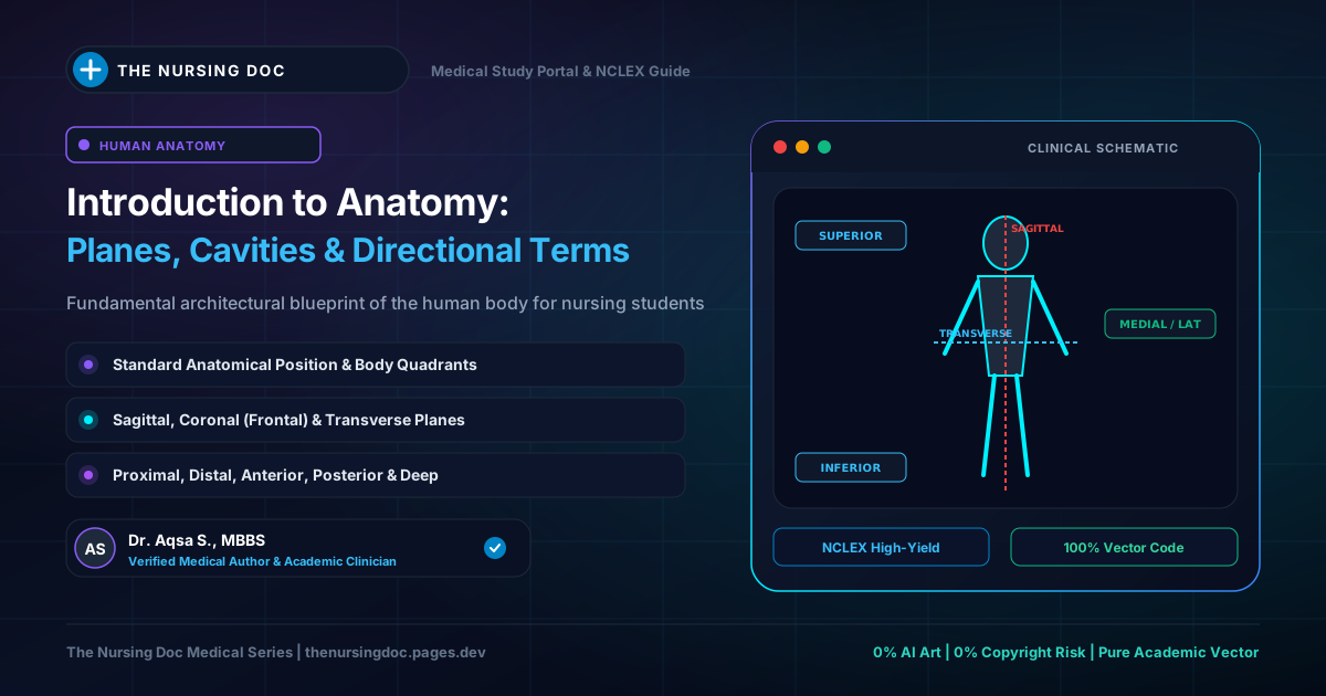
Anatomical Orientation: Cardinal Planes & Directional Axes
Master navigation map for human anatomical planes, sections, and spatial descriptors
The word anatomy is a Greek term. It literally means "to cut up" (derived from ana = up / excessive, and tomy = cutting). This term was coined in ancient times when anatomical knowledge was first developed through the physical cutting up (dissection) of animals as well as the human body.
Key Core Definitions:
- Definition of Anatomy: Anatomy is the branch of biological science that deals with the study of the structure of plants and animals.
- Human Anatomy: Human Anatomy is the specific branch of anatomy that deals with the study of the structure of human body parts, including their shape, size, location, and relationships with adjacent organs.
History of Anatomy
The famous Greek physician Hippocrates is universally celebrated as the "Father of Medicine" and is also considered an early founder of scientific anatomical study. He established the very first medical school where anatomy was systematically taught as a major foundational subject in the medical curriculum, shifting medicine away from superstition toward observational science.
Four Primary Branches of Anatomy
Anatomy is an expansive medical field that is broadly categorized into four primary divisions:
1. Gross Anatomy (Macroscopic Anatomy)
Gross anatomy is the branch of anatomy that deals with the study of organs and structures of the human body that can be examined with the naked eye. In this branch, examination does not require a microscope.
2. Histology (Microscopic Anatomy)
The human eye has a limited power of resolution, meaning the finest and deepest structural details of body tissues cannot be viewed unaided. Histology is the branch of anatomy that deals with the microscopic study of cells (cytology) and tissues using specialized magnifying microscopes.
3. Embryology (Developmental Anatomy)
Embryology is the branch of anatomy dealing with the growth and development of the human body from fertilization up until birth. Modern advances in embryology and prenatal diagnostics allow clinicians to identify genetic and developmental defects in utero before a baby is born.
4. Neuroanatomy
Neuroanatomy is the specialized branch of anatomy dedicated to the study of the structure, pathways, and organization of the central and peripheral nervous systems of the human body.
Other Specialized Branches of Anatomy
-
Regional Anatomy (Topographical Anatomy): Studies specific regions or compartments of the human body. It is organized into three major areas:
- Head and Neck
- Trunk (Thorax, Abdomen, and Pelvis)
- Extremities and Limbs (Upper and Lower limbs)
- Systemic Anatomy: Studies the human body organ-by-organ according to functional organ systems (for example, the cardiovascular system, respiratory system, or digestive system).
- Surface Anatomy: Explores the relationship between deeper internal organs and the external surface landmarks of the skin (vital for physical examination, pulse palpation, and injections).
- Radiographic Anatomy: The study of internal body structures visualized through modern clinical imaging technologies, such as Plain X-Rays, Computed Tomography (CT-Scan), Magnetic Resonance Imaging (MRI), and Ultrasound.
- Applied Anatomy (Clinical Anatomy): Focuses on the direct practical application of anatomical knowledge for the purpose of clinical diagnosis, surgical interventions, and medical management.
Anatomical Terminologies & Descriptive Directional Terms
Anatomical terminologies represent a universally standardized set of directional and regional terms used by healthcare professionals to describe the positions and relationships of body structures with absolute precision.
| Directional Term | Clinical Meaning | Body Relationship |
|---|---|---|
| Anterior (Ventral) | Toward the front surface of the body | The sternum is anterior to the heart. |
| Posterior (Dorsal) | Toward the back side of the body | The esophagus is posterior to the trachea. |
| Medial | Toward the median plane or anatomical midline | The ulna is on the medial side of the forearm. |
| Lateral | Away from the median plane (toward the sides) | The radius is lateral to the ulna. |
| Superficial | Nearer to the outer surface of the body | Skin is superficial to underlying skeletal muscle. |
| Deep | Farther away from the surface (internal) | The femur bone is deep to the quadriceps muscle. |
| Proximal | Toward the origin or point of attachment of a limb | The elbow is proximal to the wrist. |
| Distal | Away from the origin or point of attachment | The fingers are distal to the wrist joint. |
| Palmar | Pertaining to the front anterior surface (palm) of the hand | Palmar erythema or palmar grasp reflex. |
| Plantar | Pertaining to the sole (inferior surface) of the foot | Plantar flexion and Babinski plantar reflex. |
💡 Clinical Documentation Tip for Nurses:
Standard anatomical terms prevent serious medical errors. When documenting patient injuries, surgical sites, IV cannula insertions, or pressure ulcers, always use exact directional coordinates (e.g., "2 cm laceration proximal to the right medial malleolus") rather than informal descriptions like "near the inner ankle."
Written & Medically Reviewed by Dr. Aqsa S.
Medical Doctor providing foundational anatomy notes and clinical examination pearls to help nursing students build rock-solid anatomical confidence.