Protein Biochemistry: Amino Acid Dynamics, 4 Structural Tiers, & Physicochemical Classifications
An evidence-based clinical guide to protein science authored by Dr. Aqsa S. Master amino acid stereochemistry, covalent peptide bonds, the 4 hierarchical tiers of structural folding (primary to quaternary), physicochemical classes (simple, conjugated, derived), and plasma oncotic pressure homeostasis.
Curator & Medical Director, The Nursing Doc • Peer Reviewed Clinical Notes
⚡ Quick Clinical Snapshot: The Supreme Molecules of Life
- Supreme Etymology: Derived from the Greek protos ("first" or "supreme"). Proteins are nitrogenous polymers of L-alpha-amino acids linked covalently via peptide bonds.
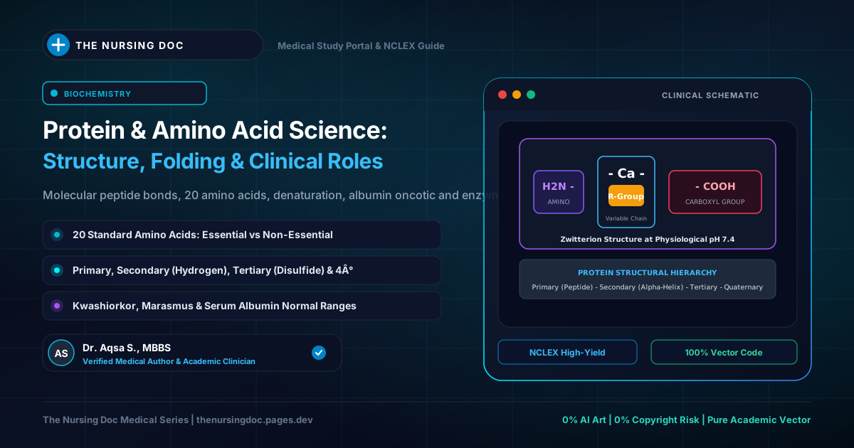
- The 20 Amino Acids: Consist of a central alpha-carbon bonded to an amino group (
-NH2), a carboxyl group (-COOH), a hydrogen atom, and a variable side chain (R-group) that determines individual chemical identity. - Four Tiers of Architecture: Primary (linear peptide-bonded amino acid sequence); Secondary (alpha-helices and beta-sheets stabilized by hydrogen bonds); Tertiary (overall 3D globular/fibrous fold driven by hydrophobic core and disulfide bonds); Quaternary (oligomeric assembly of multiple subunit chains, e.g., Hemoglobin tetramer).
- Tripartite Classification: Simple proteins (yield only amino acids: albumins, globulins, scleroproteins); Conjugated proteins (linked to a non-protein prosthetic group: lipoproteins, chromoproteins, glycoproteins); Derived proteins (denatured or hydrolytic intermediates: proteoses, peptones, peptides).
- Plasma Colloid Osmotic Pressure: Circulating plasma proteins (chiefly Serum Albumin) generate an oncotic pressure of 25–30 mmHg, retaining intravascular fluid and preventing third-space pitting edema.
From Amino Acid Monomers to Higher-Order Quaternary Macromolecules
1. Definition, Supreme Etymology, & The 20 Amino Acids
Proteins are immensely complex, high-molecular-weight nitrogenous macromolecules found ubiquitously throughout the living world. The word protein originates from the classical Greek "protos", translating directly to "the first" or "the supreme"—a testament to their indispensable preeminence in cellular architecture and physiology.
Dr. Aqsa’s Foundational Principle: "Proteins are polymers of amino acids. They are nitrogenous compounds made up of a variable number of amino acids joined to one another by a specific, rigid covalent linkage known as the peptide bond."
All natural proteins are assembled from a canonical set of 20 standard amino acids for which specific genetic codons exist in DNA and mRNA. Each standard amino acid possesses a central, tetrahedral alpha-carbon (Cα) bonded to four distinct chemical substituents:
R = H, the molecule is Glycine (H2N-CH2-COOH). Because it has two identical hydrogen atoms attached to the alpha-carbon, Glycine is the only standard amino acid that lacks a chiral center and is optically inactive.
R = CH3, the molecule is Alanine (H2N-CH(CH3)-COOH). Modifying the R-group determines polarity, charge, hydrophobicity, and specific catalytic properties across the remaining 19 amino acids.
Quantitative Distribution in Biological Systems: Proteins constitute the foundational matrix of cytoplasm and all cellular membranes without exception. Quantitatively:
- Mammalian Skeletal Muscles: 20% protein by mass.
- Blood Plasma: 7% protein (primarily albumin, globulins, fibrinogen).
- Cow's Milk: 3.5% protein (predominantly casein).
- Dietary Legumes, Beans, & Nuts: ~20% protein; Cereals: ~12% protein.
- Everyday Natural Fibers: Silk (fibroin), wool (keratin), and animal leather (collagen).
2. The Four Hierarchical Tiers of Protein Structural Organization
Every unique protein exhibits an exact, genetically dictated sequence of amino acids that spontaneously folds into a complex three-dimensional conformation essential for biological activity. Structural biologists divide this organization into four distinct tiers:
Primary Structure (Linear Polypeptide Sequence)
The fundamental linear sequence of amino acids covalently linked end-to-end via peptide linkages (-CO-NH-). By universal biochemical convention, the sequence begins at the free amino group (N-terminus on the left) and terminates at the free carboxylic group (C-terminus on the right).
Secondary Structure (Regular Spatial Folding: α-Helices & β-Sheets)
The recurring, regular spatial arrangements formed by contiguous amino acids located near each other in the linear chain. These periodic structures are stabilized strictly by hydrogen bonds formed between the carbonyl oxygen (C=O) of one peptide bond and the amide hydrogen (N-H) of another.
Tertiary Structure (Overall Three-Dimensional Conformation)
The overall three-dimensional spatial geometry assumed by an entire polypeptide chain through extensive looping, folding, and bending of secondary structural domains. Driven by non-covalent forces (hydrophobic collapse into the interior, ionic salt bridges, van der Waals forces) and covalent disulfide bridges (-S-S-) between cysteine residues.
Quaternary Structure (Multi-Subunit Oligomeric Assembly)
Not present in all proteins. Quaternary structure refers to the spatial arrangement and non-covalent association of two or more independent polypeptide chains (termed subunits) to form a single, functional, multi-subunit macromolecular complex.
3. Comprehensive Physicochemical Classification: Simple, Conjugated, & Derived
Proteins are classically categorized into three primary divisions based on their solubility, chemical composition, and physical behavior upon hydrolysis:
| Classification | Defining Biochemical Properties | Representative Sub-Classes & Examples |
|---|---|---|
| I. Simple Proteins | Yield only amino acids upon complete acid or enzymatic hydrolysis. |
• Albumins: Water-soluble, coagulable by heat (Serum albumin, ovalbumin, lactalbumin). • Globulins: Insoluble in pure water; soluble in dilute neutral salts; heat coagulable (Serum globulin, myosin, ovoglobulin). • Globins: Histidine-rich, non-basic; bind with heme to form hemoglobin. • Prolamins: Soluble in 70–80% ethanol; insoluble in water/absolute alcohol (Gliadin in wheat, Zein in maize). • Histones: Strongly basic, arginine-rich; package nuclear DNA into nucleosomes. • Protamines: Sperm cell basic proteins; lack tyrosine/tryptophan. • Albuminoids (Scleroproteins): Highly insoluble fibrous animal proteins (Collagen, Keratin, Elastin). |
| II. Conjugated Proteins | Composed of a simple protein joined covalently or non-covalently to a non-protein chemical group termed the prosthetic group. |
• Nucleoproteins: Protein + Nucleic acid (Chromatin, Ribosomes). • Phosphoproteins: Protein + Phosphoric acid (Casein in milk). • Lipoproteins: Protein + Lipids (Chylomicrons, VLDL, LDL, HDL). • Glycoproteins: Protein + Carbohydrates (<4% hexosamine: Mucins, TSH). • Chromoproteins: Protein + Pigment (Hemoglobin, Cytochromes, Rhodopsin). • Metalloproteins: Protein + Metal ion (Ferritin [Fe], Ceruloplasmin [Cu]). |
| III. Derived Proteins | Substances produced by the action of physical agents (heat, radiation), chemicals (acids, alkalis), or enzymes upon simple or conjugated proteins. |
• Primary Derived: Denatured proteins where cross-linkages are disrupted but peptide bonds remain intact (Metaproteins, Coagulated egg albumin). • Secondary Derived: Progressive hydrolytic cleavage fragments grouped by decreasing molecular weight: Proteins → Proteoses → Peptones → Polypeptides → Oligopeptides → Free Amino Acids. |
4. Systemic Biological Functions & Plasma Oncotic Homeostasis
Proteins execute an unparalleled breadth of biological duties essential for animal life:
NCLEX & Bedside Clinical Alerts
Critical pathophysiology every nurse and healthcare clinician must master
📝 NCLEX-RN Practice Check: Protein Biochemistry
Question 1: A patient presenting with decompensated liver cirrhosis has a serum albumin of 2.1 g/dL and demonstrates significant bilateral lower-extremity pitting edema and abdominal ascites. What physiological mechanism directly accounts for this fluid shift?
A) An acute surge in plasma oncotic pressure drawing interstitial fluid into lymphatic vessels.
✓ B) A reduction in plasma colloid oncotic pressure allowing intravascular fluid to extravasate into the interstitial spaces.
C) Competitive blockade of capillary endothelial sodium-potassium ATPase pumps.
D) Denaturation of erythrocyte hemoglobin quaternary structure.
Question 2: Which tier of protein structural organization describes the spatial association of two or more independent polypeptide subunits to form a single functional macromolecule, such as the heterotetramer Hemoglobin?
A) Primary structure
B) Secondary structure
C) Tertiary structure
✓ D) Quaternary structure