The Skeletal System: Bones, Cartilage, Regional Distribution, & Fracture Classifications
The 206 Bones of the Human Skeleton: Axial (80) vs. Appendicular (126)
Topographical breakdown of central axial framework and peripheral appendages
The skeletal system consists of bones and cartilage that are interconnected with each other through joints. While bones are hard, rigid, and non-flexible structures, cartilage consists of semi-rigid, tough structures that maintain significant flexibility.
Together, the skeletal framework provides our body with physical shape, structural support, mechanical movement, and essential protection for internal vital organs.
Structure of Bone Tissue:
Bones are composed of specialized osseous tissue, which is a robust variety of connective tissue. It consists of specialized bone cells (osteocytes, osteoblasts, and osteoclasts) embedded within a mineralized bone matrix (intercellular substance).
Vital Functions of Bones
The functions of bone tissue are divided into two crucial domains:
1. Mechanical Functions
- Structural Support: Bones make up the skeleton and provide the rigid framework that supports all soft tissues and maintains body posture.
- Protection: Protects vital organs like the brain (cranium), spinal cord (vertebrae), heart and lungs (rib cage), and pelvic organs.
- Movement (Levers): Bones make movement possible by serving as attachment sites for skeletal muscles and acting as levers around joints.
2. Physiological Functions
- Mineral Storehouse: Bone serves as the primary reservoir for essential minerals, particularly calcium, magnesium, and phosphate ions.
- Hematopoiesis: Bones contain red bone marrow, which serves as the primary site of blood cell production (erythrocytes, leukocytes, and platelets).
Regional Distribution of the Human Skeleton (206 Bones)
The adult human skeleton comprises 206 distinct bones, organized into two major regional anatomical divisions:
| Division | Anatomical Regions Included | Individual Bone Counts | Total |
|---|---|---|---|
| Axial Skeleton | Bones of the head, neck, and trunk along the central body axis. |
• Skull: 22 bones • Auditory Ossicles (Ears): 6 bones • Hyoid: 1 bone • Vertebral Column: 26 bones • Ribs: 24 bones (12 pairs) • Sternum: 1 bone |
80 Bones |
| Appendicular Skeleton | Bones of the upper and lower limbs, including the pectoral and pelvic girdles. |
• Upper Limbs & Pectoral Girdle: 64 bones • Lower Limbs & Pelvic Girdle: 62 bones |
126 Bones |
Classification of Bones by Shape
Bones in the human body are morphologically classified into six primary categories based on their shape, structure, and functional characteristics:
Figure 1: Morphological Shape Spectrum of Human Bones
Architectural variations matching mechanical stress and anatomical function
1. Long Bones
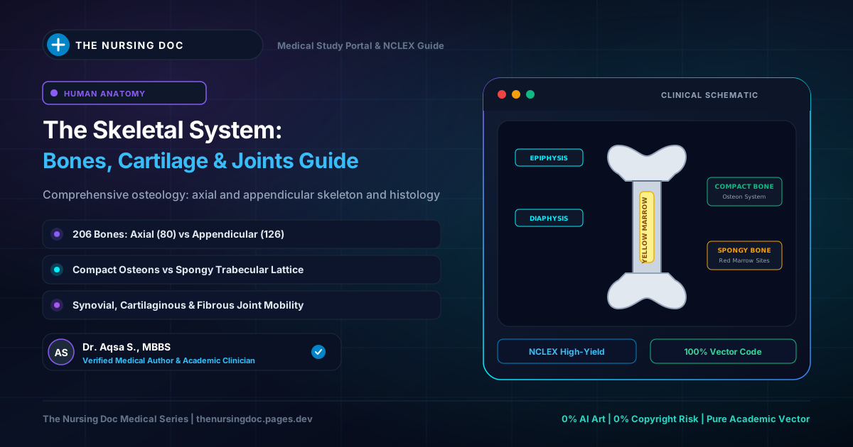
A long bone consists of a cylindrical, elongated shaft (diaphysis) and two expanded ends (epiphyses) that bear smooth articular cartilage surfaces. They are designed for weight-bearing and leverage.
Examples: Femur, Humerus, Tibia, Fibula, Radius, and Ulna.
2. Short Bones
These are generally small, roughly cube-shaped bones having nearly equal vertical and horizontal dimensions. Short bones provide localized stability with limited movement.
Examples: Carpal bones of the wrist and Tarsal bones of the foot.
3. Flat Bones
Flat bones usually occur as thin, curved plates. They serve primarily as protective coverings for delicate organs and provide broad surfaces for muscular attachments.
Examples: Parietal and Frontal bones of the cranial vault, Ribs, Sternum, and Scapulae.
4. Irregular Bones
These bones possess complex, irregular shapes with numerous bony processes that do not fit into other shape categories.
Examples: Vertebrae, Hip bones (Os coxae), and bones of the skull base and facial skeleton.
5. Sesamoid Bones
Sesamoid bones are small bony nodules embedded directly within tendons and joint capsules. They are named because their small size and contour resemble the seed of sesame plants. They protect tendons from excessive wear and alter the pull angle of muscle forces.
Examples: Patella (knee cap), pisiform bone, and small nodules in foot tendons.
6. Accessory Bones
These are secondary bones that are not uniformly present in all individuals; they may fuse with adjacent bones or remain independent. They are subdivided into:
- Supernumerary Bones: Extra digits (polydactyly) or extra ribs (cervical ribs).
- Wormian (Sutural) Bones: Small, irregular bone islands found along the cranial sutures.
- Heterotopic Bones: Bones that develop abnormally inside soft tissues as a response to repetitive trauma.
Bone Injury & Repair: Clinical Fracture Classifications
In clinical medicine, a fracture is defined as any complete or partial break in the anatomical continuity of a bone. Fractures are fundamentally categorized into two major primary classes:
Open Fracture (Compound)
A fracture that is directly associated with an external open skin wound, allowing communication between the bone fragments and the external environment.
Closed Fracture (Simple)
A fracture that produces no external wound in the overlying skin, keeping the fracture hematoma protected within intact cutaneous tissues.
🚨 Critical Nursing Alert: Open Fractures & Osteomyelitis Risk
Open (compound) fractures are orthopedic surgical emergencies! Because the protective skin barrier is violated, patients carry an exceptionally high risk of severe bacterial contamination leading to osteomyelitis (deep bone infection) and sepsis. Nurses must immediately cover open wounds with sterile saline-soaked dressings, initiate IV prophylactic antibiotics, and verify tetanus vaccination status!
Written & Medically Reviewed by Dr. Aqsa S.
Medical Doctor providing foundational anatomy, osteology, and clinical examination study notes to guide nursing students through board examinations.