Staining Techniques in Microbiology: Gram's Stain, Acid-Fast (Ziehl-Neelsen), & Diagnostic Methods
An authoritative diagnostic guide authored by Dr. Aqsa S. Master the biochemical theories of Gram's reaction, precise reagent formulations, step-by-step laboratory staining protocols, Ziehl-Neelsen staining for Mycobacterium tuberculosis, and clinical NCLEX decision-making.
Curator & Medical Director, The Nursing Doc • Peer Reviewed Clinical Notes
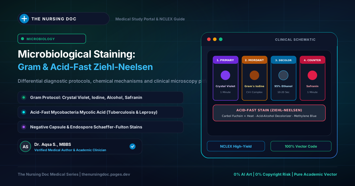
⚡ Quick Clinical Snapshot: Diagnostic Staining Principles
- Gram Staining Axiom: Gram-positive bacteria retain the primary Crystal Violet-Iodine (CV-I) complex and appear deep violet/purple due to a thick, multi-layered peptidoglycan envelope and acidic cytoplasm. Gram-negative bacteria have high lipid outer membranes that dissolve in alcohol, releasing the dye to take up Safranin counterstain and appear pink/red.
- Acid-Fast (Ziehl-Neelsen) Axiom: Mycobacterium tuberculosis possesses a dense waxy envelope rich in long-chain Mycolic Acids ($C_{60}–C_{90}$). Phenolic Carbolfuchsin penetrates with gentle steaming heat and tenaciously resists decolorization with harsh 20–25% Sulfuric Acid ($H_2SO_4$) or acid-alcohol, remaining bright magenta-red. Non-acid-fast cells counterstain light blue.
- Critical Safety Precaution: Always air-dry Acid-Fast smears vertically in a rack. Never blot dry with filter paper, as highly infectious tubercle bacilli can adhere to paper fibers, aerosolize, or cross-contaminate subsequent patient slides.
- The Decolorization Step: The single most sensitive and rate-limiting step in clinical cytology. Over-decolorization yields false Gram-negative results; under-decolorization yields false Gram-positive artifacts.
- Culture Age Factor: Gram stains must be performed on fresh cultures (18–24 hours). Autolytic enzymes in aging cultures break down the peptidoglycan sacculus, causing true Gram-positive bacteria to stain Gram-negative.
Comparative Cytological Workflows: Gram Stain vs. Ziehl-Neelsen Acid-Fast Protocol
01 Foundations of Bacterial Cytology: Why Do We Stain?
Under standard brightfield light microscopy, living bacteria are virtually transparent and invisible. The cytoplasm of a bacterial cell possesses an optical refractive index (approximately 1.33 to 1.36) that is nearly indistinguishable from that of the surrounding aqueous suspension medium or water (refractive index 1.33). Consequently, unstained bacteria pass light rays with minimal deflection or phase shift.
Staining is the biochemical process of applying synthetic organic dyes (salts composed of positive and negative ions, one of which carries the colored chromophore group) to impart contrast, enhance resolution, and delineate internal and surface morphological structures.
Simple Staining
Utilizes a single basic dye (e.g., Methylene Blue, Crystal Violet, Carbolfuchsin) applied for 30–60 seconds. Its sole purpose is highlighting general bacterial size, cellular shape (cocci, bacilli, spirilla), and cellular arrangements (clusters, chains, tetrads).
Differential Staining
Employs at least two contrasting chemical stains separated by a decolorization step (e.g., Gram's Stain and Ziehl-Neelsen Acid-Fast Stain). Segregates bacteria into distinct major taxonomic divisions based on cell wall composition.
Special / Structural Staining
Specially tailored staining protocols designed to visualize specific anatomical structures that resist standard stains, including bacterial endospores (Schaeffer-Fulton), glycocalyx capsules (Negative India ink), and flagella (Leifson).
🧪 The Biophysical Charge Mechanism of Bacterial Dyes
At physiological pH (pH 7.0–7.4), the surface and cytoplasm of bacterial cells carry a net negative electrical charge due to the abundant presence of phosphate groups in nucleic acids (DNA and RNA) and teichoic/lipoteichoic acids in Gram-positive cell walls:
- Basic Dyes (Cationic, Positive Chromophore): The colored cation ($Dye^+$) forms strong electrostatic ionic bonds with the negatively charged bacterial structures. Examples include Crystal Violet, Methylene Blue, Safranin, Malachite Green, and Basic Fuchsin. These are the workhorses of routine diagnostic microbiology.
- Acidic Dyes (Anionic, Negative Chromophore): The colored anion ($Dye^-$) is electrostatically repelled by the negative surface charge of the bacterial cell envelope. Consequently, acidic dyes do not stain the cell; instead, they stain the glass background, leaving the transparent organism illuminated as a clear halo. Examples include India Ink, Nigrosin, and Eosin.
02 Gram's Stain: Principles, Historical Theories, & Cell Wall Dynamics
Discovered empirically in 1884 by the Danish physician Hans Christian Gram while examining lung tissue from patients who died of pneumonia, the Gram stain remains the single most important and universally applied differential staining procedure in clinical medicine. It separates almost all clinically relevant eubacteria into two fundamental categories: Gram-Positive and Gram-Negative.
🔬 Three Biochemical Theories of the Gram Reaction
While modern microbiology attributes the reaction primarily to peptidoglycan thickness and membrane lipid solubility, historically three major biochemical theories were advanced to explain this remarkable phenomenon:
1. Magnesium Ribonucleate Theory
Research by Henry and Stacey demonstrated that Gram-positive organisms contain a specific surface nucleoprotein complex known as magnesium ribonucleate. If Gram-positive bacteria are stripped of magnesium ribonucleate via bile salt extraction (sodium deoxycholate) or ribonuclease digestion, they immediately lose their ability to retain crystal violet and react as Gram-negative organisms!
2. Permeability & Lipid Dissolution Theory
When iodine is added after crystal violet, an insoluble Crystal Violet-Iodine (CV-I) complex is formed. This complex is insoluble in water but soluble in organic solvents (alcohol/acetone). Gram-negative cell envelopes contain 10–20% lipids in their outer membrane; alcohol dissolves these lipids, creating large pores that allow the CV-I complex to escape easily. Gram-positive cells contain minimal lipids (1–4%) and a dense peptidoglycan mesh that dehydrates and shrinks in alcohol, sealing the CV-I complex inside.
3. Cytoplasmic pH & Acidity Theory
Gram-positive bacteria have a distinctly more acidic cytoplasm (isoelectric point around pH 2.0 to 3.0) compared to Gram-negative bacteria (pH 4.0 to 5.0). The addition of iodine acts as an oxidizing agent that further accentuates this internal cytoplasmic acidity, driving basic crystal violet to bind with tremendous electrochemical tenacity that resists alcohol elution.
Cytological Transformation of Gram-Positive vs. Gram-Negative Cells Across All 4 Steps
03 Gram Staining SOP: Step-by-Step Laboratory Protocol
Strict adherence to standard operating procedures (SOP) ensures diagnostic reliability. Deviations in timing or smear thickness lead directly to misidentification and inappropriate antimicrobial therapy.
Step 1 Smear Preparation & Air Drying
Place a small drop of sterile saline on a clean, grease-free glass slide. Using a sterile inoculating loop, emulsify a minute amount of bacterial colony to form a thin, translucent smear. Allow the slide to air dry completely at room temperature. Heating wet smears should be strictly avoided, as rapid water boiling ruptures fragile bacterial cell membranes and distorts morphology.
Step 2 Gentle Heat Fixation
Pass the completely dried slide rapidly through a Bunsen burner flame 2 to 3 times (smear side facing upwards). The slide is properly fixed when it feels comfortably warm to the dorsum (back) of your hand. Heat fixation coagulates bacterial proteins, anchoring the organisms firmly to the glass so they are not washed away during staining. Overheating chars the cell wall and induces false Gram-negativity.
Step 3 Primary Staining: Crystal Violet (1 Minute)
Place the slide horizontally on a staining rack over a sink. Flood the entire smear with 0.5% Crystal Violet (or Gentian Violet) solution and let stand for exactly 60 seconds (1 minute).
Step 4 Mordant Application: Gram's or Lugol's Iodine (1 Minute)
Tilt the slide to drain excess crystal violet. Wash off the remaining stain directly with Gram's or Lugol's Iodine solution, then flood the slide completely with fresh iodine. Leave covered for 60 seconds (1 minute). The polyiodide ions penetrate the cell envelope and react with crystal violet to precipitate the insoluble, dark CV-I mordant complex. Rinse gently with cold tap water.
Step 5 Decolorization: Acetone or 95% Ethyl Alcohol (10–20 Seconds)
Hold the slide at an angle of 45°. Add acetone or alcohol dropwise to the top of the smear until no more violet color elutes or runs from the slide (usually 5 to 15 seconds for acetone, 15 to 30 seconds for 95% ethanol). Rinse immediately with water to halt the solvent extraction action! Prolonged exposure washes the CV-I complex even out of Gram-positive cells.
Step 6 Counterstaining: Safranin / Neutral Red (30–60 Seconds)
Flood the slide with a counterstain: Safranin, Neutral Red, or 1:10 Dilute Carbolfuchsin. Leave for 30 to 60 seconds. The counterstain imparts a contrasting pink/red hue to the completely decolorized Gram-negative cells without obscuring the intense violet color of Gram-positive organisms.
Step 7 Washing, Drying, & Microscopic Examination
Rinse the slide thoroughly with cold running water. Allow the slide to air dry by placing it vertically in a draining rack, or blot gently with bibulous/filter paper (taking care not to wipe or smudge the smear). Examine under the brightfield microscope using the low-power objective (10×) to locate fields, and confirm with the 100× Oil Immersion Objective (total magnification 1000×).
04 Standard Laboratory Reagents & Chemical Formulations
Clinical quality control starts with precise reagent preparation. Below are the standard pharmaceutical laboratory formulations as utilized in medical diagnostic centers:
🟣 1. Crystal Violet Solution (0.5% Aqueous)
- • Crystal Violet powder: 0.5 g (or 5.0 g for 0.5% in 1000 mL)
- • Distilled water: 100 mL (or 1000 mL)
- • Filter through Whatman No. 1 filter paper to remove undissolved microcrystals that mimic Gram-positive cocci artifacts.
🟡 2. Lugol's / Gram's Iodine Solution
- • Elemental Iodine ($I_2$): 10 g
- • Potassium Iodide ($KI$): 20 g
- • Distilled water: 1000 mL
- • Biochemical Note: Iodine is poorly soluble in pure water; Potassium Iodide forms soluble potassium triiodide ($KI_3$), releasing reactive $I_3^-$ ions. Store in dark amber glass bottles.
⚪ 3. Decolorizing Solvents
- • Acetone-Alcohol Mixture: 500 mL Acetone + 500 mL 95% Ethyl Alcohol (rapid and balanced; standard in diagnostic labs).
- • Pure 95% Ethyl Alcohol: Slower acting, forgiving for novice technicians.
- • Pure Acetone: Ultra-rapid (decolorizes in 2–5 seconds); high risk of over-decolorization.
🔴 4. Counterstain Formulations
- • Safranin Solution: 1.7 g Safranin powder dissolved in 50 mL 95% ethanol; make up to 500 mL with distilled water.
- • Neutral Red Solution: 1.0 g Neutral Red + 2.0 mL 1% Acetic Acid + distilled water to make 1000 mL.
- • Dilute Carbolfuchsin: 1:10 dilution of Ziehl-Neelsen strong carbolfuchsin in distilled water (preferred for weakly staining organisms like Campylobacter and Bacteroides).
05 Ziehl-Neelsen (Acid-Fast) Staining: Overcoming the Mycobacterial Armor
Certain medically catastrophic bacteria—most notably Mycobacterium tuberculosis (the causative agent of tuberculosis) and Mycobacterium leprae (leprosy)—cannot be classified or stained reliably by the standard Gram technique. They frequently appear as ghost cells or unstained bacilli because their cell envelopes contain up to 60% lipid content, characterized by high-molecular-weight Mycolic Acids ($C_{60}–C_{90}$ branched beta-hydroxy fatty acids) and acyl glycolipids (such as Cord Factor).
To overcome this waxy, hydrophobic impermeability barrier, German bacteriologists Franz Ziehl and Friedrich Neelsen developed the Acid-Fast Staining technique in 1882–1883.
🛡️ The Biophysical Principle of "Acid-Fastness"
The term Acid-Fast designates the unique cytological property of an organism to tenaciously resist decolorization by strong mineral acids ($20–25\% \text{ H}_2\text{SO}_4$) and alcohol once stained by a lipid-soluble dye:
- The Carrier Solvent (Phenol): Basic Fuchsin is dissolved in a high concentration of aqueous phenol (forming Carbolfuchsin). Phenol is lipophilic and acts as a solvent vehicle, dissolving into the lipid-rich mycolic acid barrier.
- The Thermal Penetrant (Steaming Heat): Intermittent gentle flame heating underneath the slide liquefies the waxy mycolic waxes, allowing carbolfuchsin to penetrate deep into the bacterial cytoplasm and bind to ribosomal RNA.
- Trapping the Dye: Upon cooling, the waxy envelope re-solidifies, effectively trapping the carbolfuchsin. Even aggressive decolorization with 25% sulfuric acid or 3% hydrochloric acid in alcohol fails to extract the dye from the waxy matrix.
- Microscopic Contrast: Organisms lacking mycolic acids (non-acid-fast) are rapidly stripped of carbolfuchsin by the acid decolorizer and take up the counterstain (Methylene Blue), creating a vivid diagnostic contrast under the microscope.
Mycolic Acid Barrier vs. Acid Decolorization Resistance
06 Ziehl-Neelsen Clinical Procedure & Mandatory Biohazard Safety
Diagnosis of active pulmonary tuberculosis relies heavily on the direct microscopic examination of sputum smears stained by the Ziehl-Neelsen method. The step-by-step SOP and critical safety rules are detailed below:
1 Smear Preparation from Clinical Specimen
Select a purulent or blood-tinged portion of early morning sputum using a sterile wooden applicator stick or wire loop. Spread an even, 2 × 1 cm oval smear on a labeled glass slide. Allow to dry completely at room temperature, then heat-fix by passing through a flame 3 to 4 times.
2 Carbolfuchsin Flooding & Intermittent Steaming Heat
Flood the entire slide with strong Ziehl's Carbolfuchsin (basic fuchsin + 5% phenol). Using a spirit lamp or Bunsen flame, heat gently underneath the slide until steam rises visibly. Keep the slide steaming for 5 minutes. Do NOT allow the stain to boil or dry out; if the stain evaporates, add additional drops of carbolfuchsin immediately. Rinse gently with cold tap water.
3 Decolorization with 20–25% Sulfuric Acid ($H_2SO_4$)
Flood the slide with 20% to 25% Sulfuric Acid ($H_2SO_4$) or acid-alcohol (3% concentrated $HCl$ in 95% ethanol). Leave the acid on the slide for 1 to 2 minutes, wash with water, and repeat until the smear appears faintly pink or pale grey. Rinse thoroughly with water, then pour on 95% ethyl alcohol for 2 minutes to extract any remaining free red dye from the background.
4 Counterstaining with Methylene Blue (1–2 Minutes)
Flood the slide with Loeffler's Methylene Blue (or 0.5% Malachite Green / Picric Acid) for 1 to 2 minutes. Rinse thoroughly with water.
⚠️ Mandatory Clinical Safety Precaution: Vertical Drying Rule
DO NOT BLOT DRY WITH FILTER PAPER! Unlike routine Gram smears, Acid-Fast slides must always be placed vertically in a drying rack and allowed to air-dry naturally.
The Clinical Hazard: Mycobacterium tuberculosis organisms possess an exceptionally sticky, waxy surface coat. If a technician blots an AFB smear with bibulous paper, viable or dead tubercle bacilli readily adhere to the porous paper fibers. If that same filter paper is subsequently used to blot another patient's slide, bacilli will transfer, producing catastrophic false-positive tuberculosis reports and creating an occupational airborne biohazard for laboratory staff!
🔬 Microscopic Interpretation & Morphology Under Oil Immersion (1000×)
Acid-Fast Bacilli (AFB Positive):
Appear as bright magenta-red, slender, slightly curved or beaded rods (0.5 to 3 μm in length). They frequently appear singly, in small clusters, or arranged in parallel serpentine cords due to Trehalose 6,6'-dimycolate (Cord Factor).
Non-Acid-Fast Background:
All non-acid-fast bacteria, respiratory epithelial cells, polymorphonuclear neutrophils (pus cells), and mucous debris take up the counterstain and appear pale blue, providing an ideal contrasting background.
07 Special & Ancillary Diagnostic Staining Techniques
Beyond Gram and Ziehl-Neelsen, medical microbiologists utilize specialized cytological stains to reveal unique anatomical structures essential for definitive microbial identification:
| Staining Technique | Target Organism / Structure | Key Reagents | Diagnostic Appearance |
|---|---|---|---|
| Schaeffer-Fulton Endospore Stain | Bacterial Endospores (Bacillus anthracis, Clostridium tetani) | Malachite Green (heated with steam) + Safranin counterstain | Bright green oval endospores within pink/red vegetative bacilli. |
| Negative / India Ink Capsule Stain | Polysaccharide Capsules (Cryptococcus neoformans, K. pneumoniae) | India ink or 10% Nigrosin (acidic background stains) | Clear refractive luminous halos surrounding yeast cells against pitch-black background. |
| Albert's Stain | Volutin / Metachromatic Granules of Corynebacterium diphtheriae | Albert's I (Toluidine blue, Malachite green, Glacial acetic acid) + Albert's II (Iodine) | Bluish-black beaded granules located at the poles of green rod-shaped bacilli. |
| Fontana's Silver Impregnation | Ultrathin Spirochetes (Treponema pallidum, causative agent of Syphilis) | Silver nitrate and ammoniacal silver developer solutions | Silver precipitates onto submicroscopic flagella/cell walls, turning delicate spirals thick jet-black. |
08 Clinical Troubleshooting: Common Laboratory Errors & Pitfalls
In diagnostic microbiology, technical errors in slide preparation lead to incorrect Gram categorizations, which can misguide empiric antibiotic choices (e.g., treating with Vancomycin when Ceftriaxone is required). Watch out for these 5 high-yield pitfalls:
1. Over-Decolorization (The #1 Lab Error)
Leaving acetone or alcohol on the slide for more than 20 seconds leaches the CV-I complex even through thick peptidoglycan layers. Result: Gram-positive cocci (e.g., Staphylococcus aureus) falsely appear as Gram-negative pink cocci!
2. Under-Decolorization
Inadequate washing with alcohol fails to extract the CV-I complex from the thin lipid outer membranes of Gram-negative bacteria. Result: Gram-negative bacilli (e.g., E. coli) falsely retain purple dye and are misdiagnosed as Gram-positive rods!
3. Aging Culture Autolysis (>24 Hours Old)
As bacterial cultures age past 24 hours, autolytic enzymes digest the peptidoglycan cell wall. The wall becomes porous, unable to retain crystal violet. Always perform Gram stains on fresh, actively growing colonies (18 to 24 hours).
4. Excessive Heat Fixation
Holding the slide in the flame too long scorches the bacterial envelopes, rupturing cell walls and destroying antigenicity. The bacteria lose stain retention and lyse into unidentifiable cellular debris.
🩺 NCLEX & Medical Board Clinical Pearls: Cerebrospinal Fluid (CSF) Gram Stains
The Stat CSF Gram Stain: When acute bacterial meningitis is suspected, a lumbar puncture is performed immediately. The CSF is centrifuged and an urgent stat Gram stain is prepared.
- Gram-Negative Diplococci (Kidney bean shaped): Diagnostic of Neisseria meningitidis (Meningococcus). Requires immediate IV Ceftriaxone and respiratory droplet isolation!
- Gram-Positive Diplococci (Lancet shaped): Diagnostic of Streptococcus pneumoniae (Pneumococcus). Requires immediate Vancomycin + Ceftriaxone + Dexamethasone.
- Gram-Positive Bacilli: Suspect Listeria monocytogenes in neonates, elderly, or immunocompromised patients. Requires IV Ampicillin!
📝 High-Yield NCLEX & Medical Board Review Questions
Test your diagnostic reasoning on microbiological staining techniques and clinical interpretation.
Question 1: A laboratory technician performs a Gram stain on a known culture of Staphylococcus aureus. Upon microscopic examination, the cocci appear bright pink instead of violet. Which procedural error is the most probable cause of this finding?
A) The technician forgot to apply the safranin counterstain.
B) The crystal violet was left on the slide for 2 minutes instead of 1 minute.
✓ C) Excessive decolorization with acetone-alcohol leached the crystal violet-iodine complex from the thick peptidoglycan wall.
D) The smear was allowed to air dry at room temperature before gentle heat fixation.
Question 2: While preparing an Acid-Fast (Ziehl-Neelsen) smear from an inpatient with suspected cavitary pulmonary tuberculosis, why is the technician strictly instructed to air-dry the slide vertically rather than blotting with bibulous paper?
A) Blotting paper dissolves the carbolfuchsin stain from the cell wall.
✓ B) Tubercle bacilli can adhere to the porous paper fibers, causing biohazard exposure and cross-contaminating subsequent patient slides with false-positive results.
C) Blotting paper creates an alkaline reaction that destroys mycolic acid bonds.
D) Filter paper fibers mimic Gram-negative bacilli under brightfield illumination.
Question 3: In the Ziehl-Neelsen acid-fast staining procedure, which cellular component is responsible for the organism's ability to resist decolorization by 25% sulfuric acid?
A) High concentration of teichoic and lipoteichoic acids.
B) A dense multi-layered murein peptidoglycan sacculus.
✓ C) Long-chain beta-hydroxy fatty acids known as Mycolic Acids ($C_{60}–C_{90}$).
D) Outer membrane lipopolysaccharide (LPS) lipid A endotoxin.