Tissue Physiology: The 4 Primary Tissues, Connective Matrix, & Epithelial Classification
⚡ High-Yield Clinical Summary
- Tissue Definition: An organized aggregation of specialized cells and associated intercellular matrix coordinated to perform specific physiological duties.
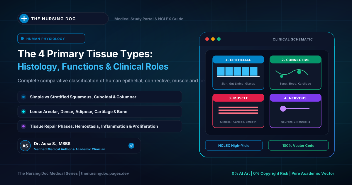
- 4 Cardinal Tissues: Epithelial Tissue, Connective Tissue, Muscular Tissue, and Nervous Tissue.
- Connective vs. Epithelial Rule: Connective tissue features few cells suspended in an abundant extracellular matrix. Epithelium features tightly packed cells with minimal intercellular substance.
- Connective Matrix Components: Extracellular fibers (Collagen, Reticular, Elastic) plus Ground Substance (Proteoglycans & Glycoproteins).
- Epithelial Categories: Simple (single layer, absorption & secretion) vs. Stratified (multiple layers, mechanical & chemical protection).
The 4 Primary Tissues of the Human Body: Architectural Spectrum
Cellular density, extracellular matrix abundance, and specialized bodily roles
1. Definition & The Four Basic Body Tissues
In human physiology and histology, a tissue is defined as a collection of specialized cells and their associated intercellular material (extracellular substance) working harmoniously to execute one or more specific bodily functions.
All organs and structures throughout the human body are constructed from combinations of four fundamental primary tissue families:
- 1. Epithelial Tissue (Epithelium): Protective sheets lining external body surfaces, internal organ cavities, and glandular secretory units.
- 2. Connective Tissue: Structural, metabolic, and immune framework binding and supporting bodily structures.
- 3. Muscular Tissue: Excitable, contractile fibers (Skeletal, Cardiac, Smooth) responsible for voluntary and involuntary movement.
- 4. Nervous Tissue: Electrically excitable neurons and supporting neuroglia coordinating neuro-endocrine bodily communication.
Epithelial vs. Connective Tissue: Architectural Differences
| Histologic Parameter | Epithelial Tissue | Connective Tissue |
|---|---|---|
| Cell Density | Extremely dense, closely packed cells forming continuous barriers. | Relatively few, widely dispersed cells. |
| Intercellular Matrix | Minimal / scant intercellular material between cells. | Large, dominant amount of extracellular substance (fibers + ground substance). |
| Vascularity | Avascular (depends on diffusion from underlying vascular bed). | Typically highly vascularized (except cartilage and dense regular tendons). |
| Basement Membrane | Always rests upon a basal lamina / basement membrane. | Underlies basement membranes; supports adjoining tissues. |
| Primary Roles | Protection, absorption, secretion, filtration. | Support, binding, nutrient transport, immune surveillance, lipid storage. |
2. Connective Tissue Architecture
Connective tissue is distinctly characterized by having relatively few cells embedded within an expansive, non-living intercellular substance (matrix).
Fundamentally, all varieties of connective tissue are constructed from three core components:
- Cells: The living functional units synthesizing and maintaining the extracellular environment.
- Fibers: Structural protein threads providing tensile strength and elastic recoil.
- Ground Substance: An amorphous, hydrated gel embedding fibers and cells.
💡 Histology Rule: Fibers + Ground Substance = Matrix (Intercellular Substance). A clear description of these matrix components is necessary before approaching the broader classification of connective tissues.
Figure 1: The Connective Tissue Triad (Areolar Tissue Prototype)
Cellular population suspended in protein fibers and proteoglycan ground substance
Types of Connective Tissue Cells
Connective tissue contains diverse cell populations. Importantly, loose areolar connective tissue acts as the premier histologic model because almost all varieties of connective tissue cells can be observed within it:
- • Fibroblasts: The primary and most abundant resident cells. They synthesize the extracellular matrix components including collagen, reticular, and elastic fibers, along with ground substance proteoglycans.
- • Histiocytes (Tissue Macrophages): Phagocytic immune cells derived from monocytes that patrol connective tissue, engulfing cellular debris, foreign microorganisms, and particulate matter.
- • Plasma Cells: Effector B-lymphocytes with characteristic clock-face nuclear chromatin. They are dedicated antibody factories that actively synthesize and secrete immunoglobulins into interstitial fluid.
- • Mast Cells: Perivascular immune sentinels loaded with basophilic granules containing histamine (mediator of vasodilation and allergic symptoms) and heparin (anticoagulant).
- • Fat Cells (Adipose Cells / Adipocytes): Cells specialized for lipid storage. They contain a massive central triglyceride droplet that compresses the nucleus against the periphery, providing thermal insulation, protection, and metabolic energy storage.
- • Wandering Cells: Occasional visitors from the circulating blood (such as neutrophils, eosinophils, and monocytes) that migrate into the connective matrix during tissue injury or immune responses.
Connective Tissue Fibers
Connective tissue fibers are classified into three distinct physical categories. The relative proportion of these fibers varies across different connective tissue subtypes:
1. Collagenous Fibers
Composed of collagen protein. Inextensible, flexible, and endowed with tremendous tensile strength to resist mechanical traction (e.g., in tendons, fascia, and organ capsules).
2. Reticular Fibers
Extremely fine, branching fibers forming delicate, mesh-like structural networks (stroma) that cradle cells in lymphoid organs (lymph nodes, spleen, red bone marrow).
3. Elastic Fibers
Formed by elastin protein and microfibrils. Capable of significant stretching and spontaneous recoil back to original dimensions without tissue damage (found in pulmonary tissue, vocal cords, and elastic arteries).
Ground Substance of the Connective Tissue
The ground substance is composed mainly of macromolecular protein-polysaccharide complexes known as proteoglycans. In addition, it contains structural glycoproteins, water, and dissolved electrolytes:
-
Proteoglycans: Large complexes responsible for binding water and creating hydrated viscoelastic gels. Diverse varieties of proteoglycans and glycosaminoglycans are distributed throughout bodily tissues, including:
- Hyaluronic Acid: Abundant, lubricating non-sulfated GAG in synovial joints, skin, and vitreous humor.
- Chondroitin Sulfate: Major structural component providing compression resistance in cartilage and bone.
- Dermatan Sulfate: Present in skin, tendons, blood vessels, and heart valves.
- Keratan Sulfate: Key hydration proteoglycan in the cornea and cartilage.
- Heparan Sulfate: Regulates cellular adhesion and vascular permeability.
- Glycoproteins: Like proteoglycans, glycoproteins are composed of proteins and carbohydrates. However, in contrast to proteoglycans, the protein moiety predominates in glycoproteins. Examples like fibronectin and laminin act as structural anchors holding cells to extracellular collagen.
3. Epithelial Tissues: Characteristics & Types
Epithelial tissues form the continuous covering of external body surfaces, line internal body cavities, tubes, and organ lumens, and constitute secretory glands.
The histological structure of epithelium is intimately coupled to its core physiological functions:
🛡️ 1. Protection
Protects underlying vulnerable structures from chemical abrasion, mechanical injury, bacterial invasion, and lethal water loss (dehydration).
🧪 2. Secretion
Specialized epithelial cells release mucus, enzymes, perspiration, and hormones from internal and external glands.
🧽 3. Absorption
Single-layer epithelial linings in the digestive tract and renal tubules selectively absorb water, electrolytes, and vital nutrients.
Epithelial cells are very closely packed together, and the intercellular substance (matrix) is minimal.
Classification: Simple vs. Stratified Epithelium
Based upon cell layer architecture, epithelial tissues are divided into two fundamental categories:
1. Simple Epithelial Tissues (Single Layer)
Simple epithelium consists of a single layer of identical cells resting directly on the basement membrane. It is primarily situated on absorptive or secretory surfaces where rapid diffusion, filtration, or secretion is needed. Simple epithelium is divided into four major varieties:
- Simple Squamous: Single layer of flattened, scale-like cells (e.g., lining of pulmonary alveoli and capillary walls).
- Simple Cuboidal: Single layer of cube-shaped cells specialized for secretion and absorption (e.g., kidney tubules, salivary ducts).
- Simple Columnar: Single layer of tall, column-shaped cells (e.g., lining of stomach and small intestine).
- Ciliated / Pseudostratified Columnar: Columnar cells equipped with surface cilia (e.g., lining the upper respiratory tract to sweep mucus).
2. Stratified Epithelial Tissues (Multilayered)
Stratified epithelia consist of several layers of cells of various shapes. The main function of stratified epithelium is to protect underlying structures against mechanical wear, friction, and environmental stresses:
- Stratified Squamous (Keratinized): Dry, tough, water-resistant outer layer of the skin (epidermis).
- Stratified Squamous (Non-Keratinized): Moist protective lining subject to abrasion (oral cavity, pharynx, esophagus, vagina).
- Transitional Epithelium: Specialized stratified cells that stretch and flatten under fluid tension (lining urinary bladder and ureters).
Figure 2: Cellular Morphology & Stratification of Epithelial Tissues
Single-layer absorptive/secretory sheets vs. multi-layer friction shields
⚠️ NCLEX & Clinical Pearls for Nurses
- Anaphylaxis & Mast Cells: Mast cells release massive amounts of histamine during Type I hypersensitivity reactions. Nurses must anticipate immediate administration of Intramuscular (IM) Epinephrine to reverse life-threatening hypotension and laryngeal edema.
- Plasma Cells & Multiple Myeloma: Neoplastic transformation of plasma cells leads to multiple myeloma, characterized by bone pain, hypercalcemia, renal failure, and elevated serum monoclonal (M) protein.
- Wound Repair & Fibroblasts: In the proliferative phase of wound healing, fibroblasts synthesize collagen fibers. Protein malnutrition and Vitamin C deficiency prevent proper collagen synthesis, leading to surgical wound dehiscence.
- Carcinoma vs. Sarcoma: Malignancies arising from epithelial tissue (due to frequent cell division) are called carcinomas (e.g., breast, lung, colon cancer). Malignancies arising from connective tissue are called sarcomas (e.g., osteosarcoma, liposarcoma).
Written & Medically Reviewed by Dr. Aqsa S.
Medical Doctor curating foundational histology, anatomy, and physiology study guides to empower nursing students with clinical precision and NCLEX confidence.